Echoes Issue No. 30 [ December 2022 ]

Welcome to the latest edition of ECHOES ....
As 2022 draws to a close we reflect on a very successful WFUMB conference in Timisoara in Romania, the launch of Ultrasound Connections, more societies joining the WFUMB family and a rapidly increasing website usage rate. It is also with much sadness that we announce the passing of two of WFUMB's past councillors. Dr Beryl Benacerraf was WFUMB treasurer from 2003-2011 and Dr Marvin Ziskin, WFUMB President 2003-2006, who gave a Luminary interview for our March 2022 edition of ECHOES
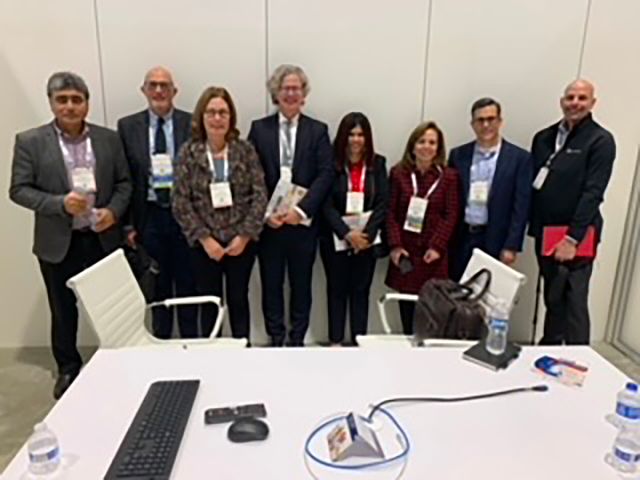
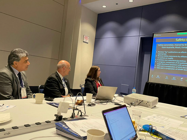
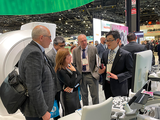
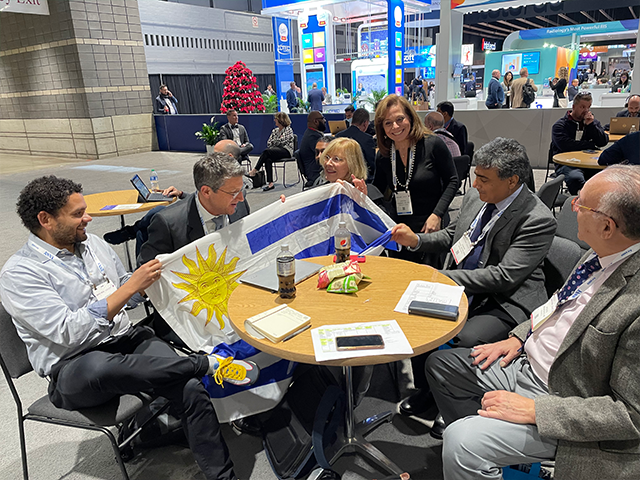
In this edition of ECHOES we have reports from CoE events in Fiji, Uganda, Indonesia and Peru, the team's visit to RSNA 2022, the Cambodian Midwife Project, a WFUMB supported course in Zanzibar, a Luminary Interview with David H Evans, updates from WFUMB 2023 and much more!
Please help us spread the word that WFUMB is there for all users of ultrasound by passing on the link to the WFUMB website & ECHOES to all your colleagues.
Have a safe and happy festive season and may 2023 bring us together to share more ultrasound education.
Sue Westerway,
Publications Committee
Intelligent Ultrasound to support WFUMB in improving global healthcare in underserved areas of the world
Intelligent Ultrasound is delighted to be supporting the World Federation for Ultrasound in Medicine and Biology (WFUMB) in its mission to bring sustainable ultrasound programs to the underserved areas of the world to improve global healthcare through collaboration, communication and education.
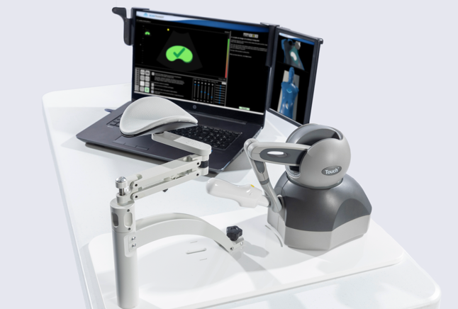
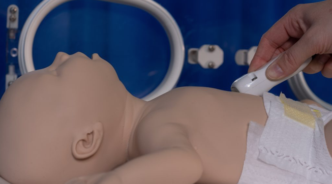
The partnership will see Intelligent Ultrasound donate a number of its world leading ultrasound training simulators to WFUMB to support their ongoing ultrasound education programme.
The simulators include the ScanTrainer Compact obstetrics and gynaecology ultrasound simulator and the new BabyWorks neonate and paediatric training simulator that was launched at the beginning of this year.
Intelligent Ultrasound will also provide training for WFUMBs congresses and regional Centre of Education courses using its new web-based training facility in Cardiff.
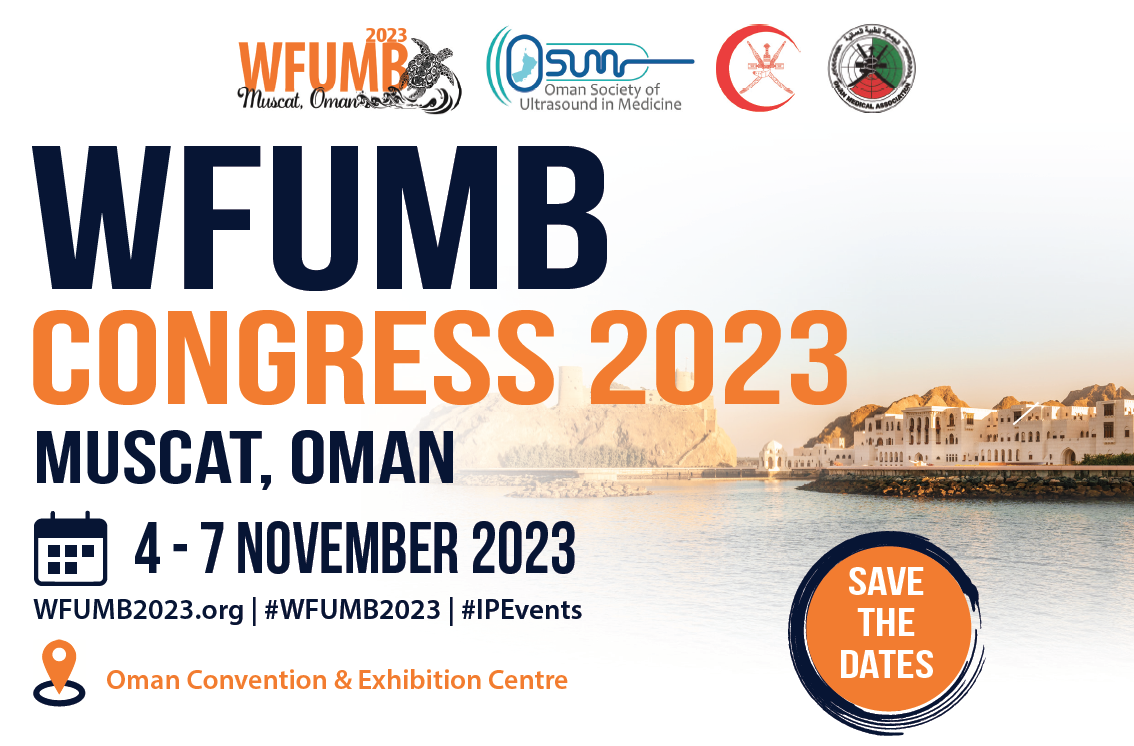
Two of the key WFUMB events in 2023 will be the education courses that will be held at EUROSON 2023 in Riga, Latvia in May and the WFUMB Congress to be held in Muscat, Oman in November.
Cristina Chammas, President and Jacques Abramowicz, President-Elect of WFUMB said
“This amazing donation of a ScanTrainer and BabyWorks for obstetrics, gynaecology and paediatric ultrasound members and trainees will provide invaluable hands-on experience at our worldwide Centres of Education courses and congresses.”
Stuart Gall, CEO of Intelligent Ultrasound said
“We are delighted to be working with WFUMB to support their ongoing mission to bring sustainable ultrasound programs to the underserved areas of the world. Education is a key part of this programme and the ScanTrainer and BabyWorks simulators are simulation at its best, providing very realistic practice in necessary and potentially lifesaving skills that are otherwise quite difficult to teach at the bedside. We look forward to working with WFUMB and supporting them in their excellent education programmes.”

Ultrasound in Medicine & Biology
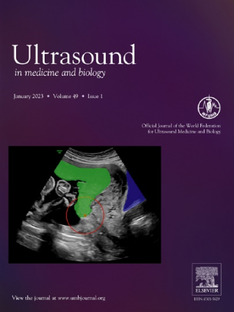
WFUMB is pleased to announce that in 2022 starting with VoI 48, Issue 11 there are new style changes to the journal
- A fresh and exiting Front Cover with changing image reflecting journal’s content in every issue
- Font change and modern manuscript layout from Vol 49, Issue 4
- Reference format using Vancouver format with numbering
- Mast Head Changes
- Shown History dates
- Updated Instructions for Authors with revised article types
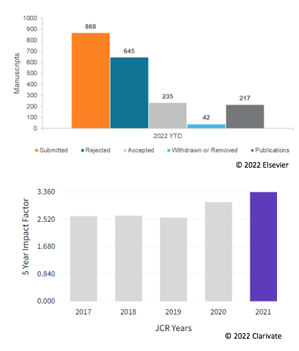
Submitted Manuscripts & Editorial Outcomes in 2022
All time High Impact Factor in 2021 (3.694)
Learn more about our journal and submit your research today!
Top Cites Articles
- Guidelines and Good Clinical Practice Recommendations for Contrast-Enhanced Ultrasound (CEUS) in the Liver–Update 2020 WFUMB in Cooperation with EFSUMB, AFSUMB, AIUM, and FLAUS. Christoph F. Dietrich et al., October 2020
- Super-resolution Ultrasound Imaging. Kirsten Christensen-Jeffries et al., April 2020
- Ultrasound-Responsive Cavitation Nuclei for Therapy and Drug Delivery. Klazina Kooiman et al., June 2020

WFUMB's Open Access Journal Launch


Latest Partnerships and MoU's
The WFUMB council has been working hard to foster new Partnerships and create new Memorandums of Understanding between WFUMB and the following organisations ...

RAD-AID is a nonprofit public service charity organization (tax-exempt under section 501(c)(3) of the United States Internal Revenue Code) helping medically underserved communities and low-resource institutions throughout the world to increase and improve radiological and medical imaging health care capabilities.
RAD-AID International (“RAD-AID”) and The World Federation for Ultrasound in Medicine and Biology (WFUMB), are collaborating for the joint purpose of advancing ultrasound in low-resource regions of the world.
To view the RAD-AID website visit: https://rad-aid.org/

The International Federation of Medical Students Associations (IFMSA) is one of the world’s oldest and largest student-run organisations. IFMSA was established in 1951 after the end of World War II to serve as a platform for unity, collaboration and peace among medical students.
It represents, connects and engages every day with an inspiring and engaging network of 1.3 million medical students from 130 countries worldwide and working in close relation with several non-governmental organisations from all over the world.
IFMSA and WFUMB intend to create joint capacity-building activities taking place either virtually or physically that will benefit members of both organisation. Both parties will collaborate on research efforts on the topics relevant to their work, such as student ultrasound education (e.g., providing input and increasing outreach.).
View the IFMSA website here https://ifmsa.org/

Imaging the World works to improve people’s lives by increasing access to modern medical imaging technology in the most rural and resource-limited areas of the world.
WFUMB looks forward to collaborating with ITW.
To view the ITW website visit: https://www.imagingtheworld.org/
World Medical Colours (WMC) has been active in Zanzibar Archipelago since several years, and the results obtained with the first courses have been published in the AJR journal in 2007 (doi: 10.2214/AJR.07.2202). The WMC ultrasound training program has grown over the years and since 2011 there is a signed agreement with the Ministry of Health.
WMC and WFUMB aim to collaborate on global projects including website content, research, training methods, sponsored mentoring and contributions in the WFUMB UMB and Open Access Journal.

The World Federation of Pediatric Imaging (WFPI) provides an international platform for pediatric radiology organizations united to address the challenges in global pediatric imaging training and the delivery of services.
WFUMB looks forward to collaborating with WFPI on future projects.
To view the WFPI website visit: https://www.wfpiweb.org/

Ultrasound course in Kampala, Uganda October 2022
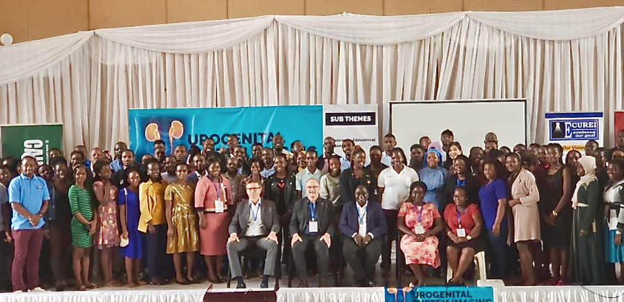
The chairman of WFUMB CoE in Uganda Prof. Michael Kawooya organized a post-graduate ultrasound course in Kampala with UGASON and ECUREI. WFUMB was represented at the course with Prof. Dieter Nurnberg and Chair of CoE TFG Prof. Odd Helge Gilja. We also arranged hands-on sessions with elastography of the liver.
During 4 conference days (Students Meeting, Preconference, Conference) from 19th to 22nd October the main topics were: Abdominal and urogenital imaging and new methods and challenges. They gave lectures (no. 10) about new methods (CEUS and elastography) and urogenital topics and interventional US. At the last day local speakers presented fine results of research in AI (artificial intelligence). 80 to 160 people attend the meeting every day to learn and discuss with the international lecturers. Students, sonographers and radiologists from Uganda and neighboring countries participated. The famous Makerere University, 100 years annversary 2022, was an appropriate location for the successful Conference. Different sponsors supported the meeting by exhibitions and hands-on training at the Mengo Hospital/ECUREI. Especially in elastography the participants were very interested. UGASON and Michael Kawooya was very generous hosts and responsible for a highly interesting and well organized Conference. It was impressive to observe the great work of Kawooya and his tream in the WFUMB COE and ECUREI institute promoting Ultrasound education in Uganda in an excellent way.
ZNZ, 1/11 -2022,
Prof. Dieter Nurnberg and Prof. Odd Helge Gilja
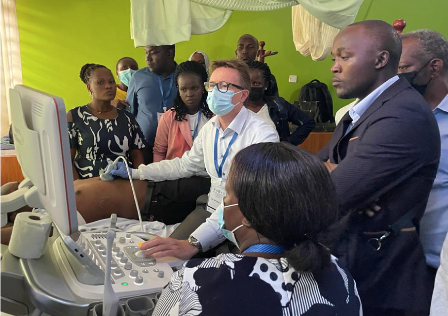
Prof. Gilja is demonstrating elastography, a new topic for the participants and of high interest
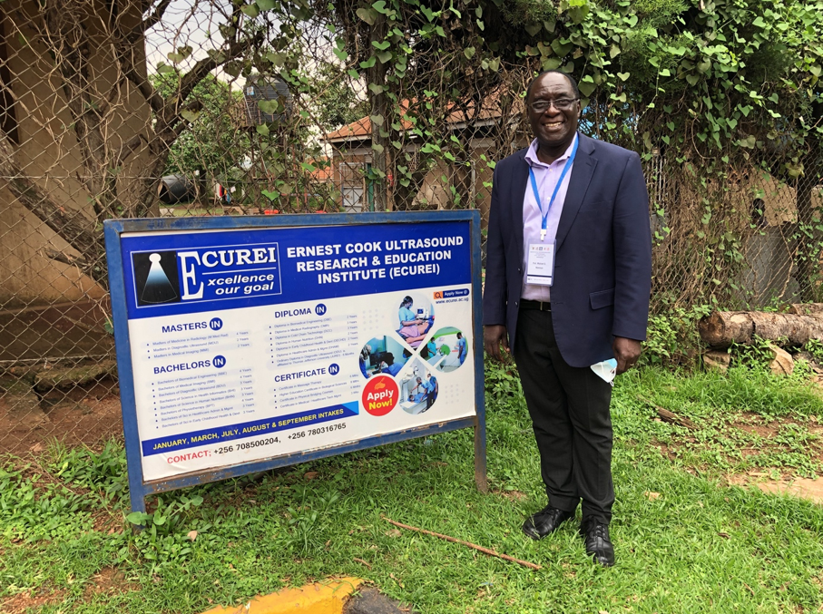
Prof Michael Kawooya at the WFUMB CoE in the ECUREI Institute
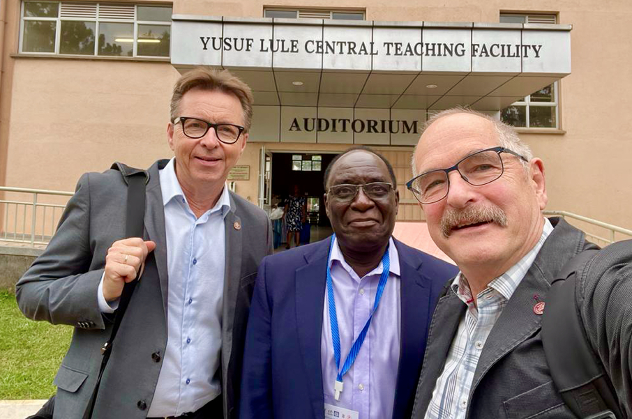
Prof. Kawooya in the middle of the international faculty

Visiting Professor Program, Zanzibar November 2022
I was appointed by the ExB to attend as a visiting professor the ultrasound course organized by Dr. Odd Helge Gilja through the University of Bergen-Norway, which was held in Zanzibar-Tanzania, from October 31 to November 3, 2022.
The lectures I delivered were:
- Physical Principles of Ultrasound and Image Optimization
- Doppler: methodology and techniques
- Doppler Key Applications: Kidney and Carotids
- POCUS Doppler: AAA, DVT, Vena Cava.
- POCUS: need in developing countries
- FAST, EFAST, BLUE and FATE Protocols
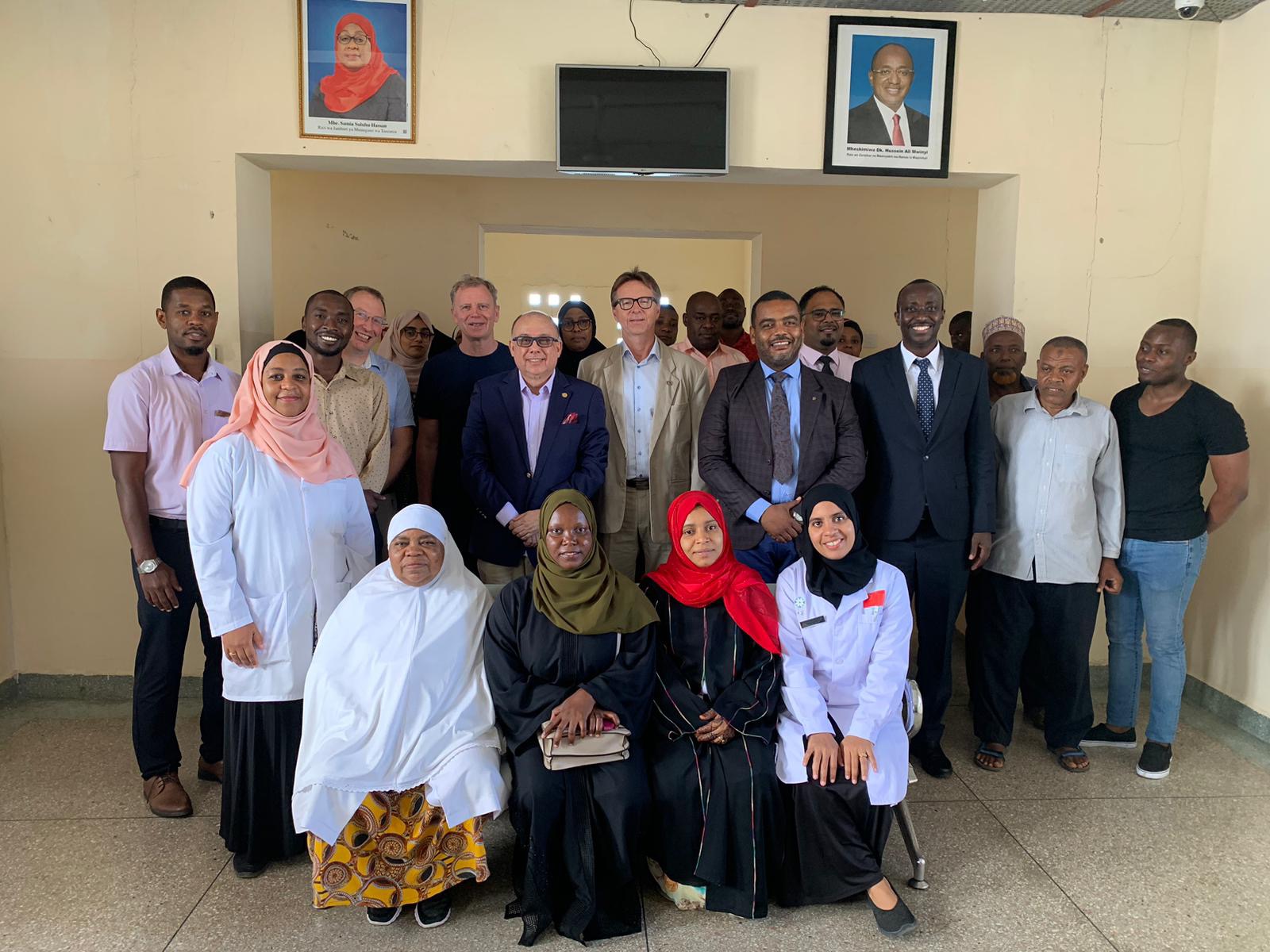
The course was well organized and held at the Mnazi Mmoja Hospital in Stone Town, in an appropriate room with good projection system, Smart TV, WiFi and air conditioning.
We had 25 participants between technicians and doctors, some with very basic ultrasound management and others with more experience and skill. Everyone showed great interest on the lectures.
Dr. Gilja and two other Norwegian professors presented topics of interest to the audience, with a focus mainly on the area of gastroenterology. They also presented lectures on CEUS and hepatic elastography.
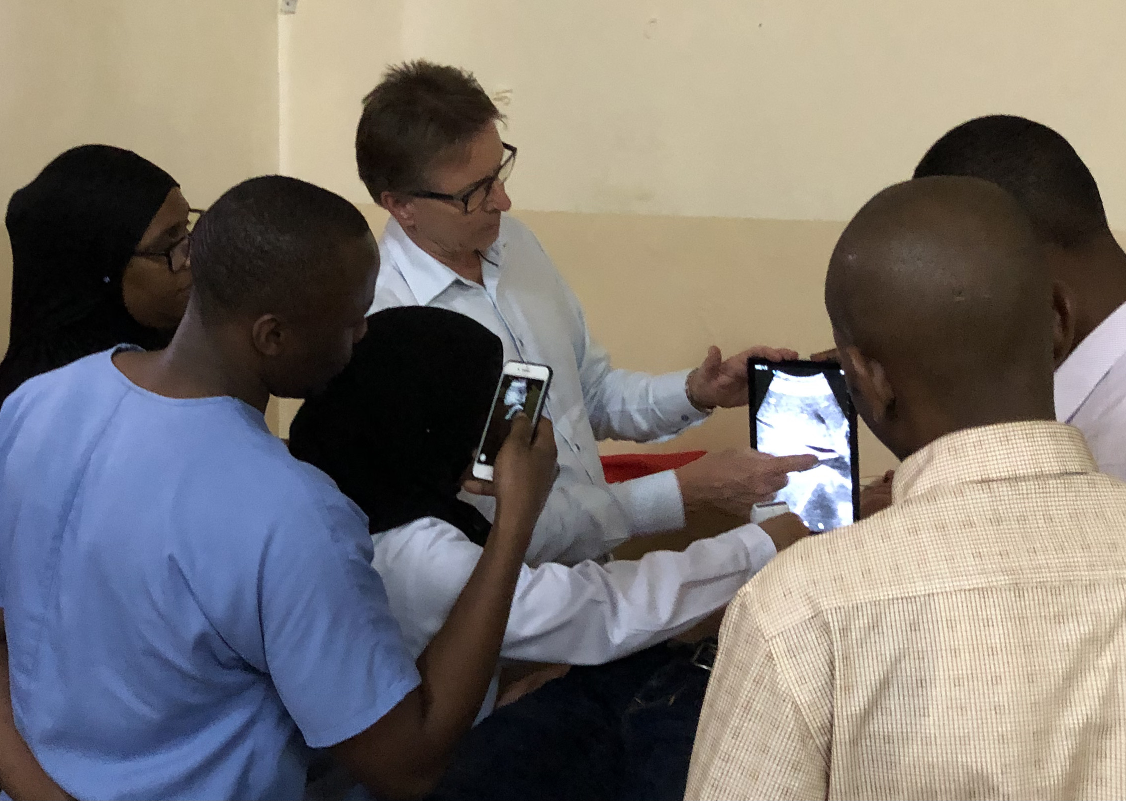
For the practical sessions all the speakers brought our ultra-portable devices. In my case, I used the Butterfly system assigned to the WFUMB COE Venezuela.
![Echoes Issue No. 30 [ December 2022 ]](https://wfumb.info/wp-content/uploads/2022/12/IMG_1437-scaled.jpeg)
The hospital authorities showed up on the first and last day of the event and thanked the University of Bergen and WFUMB for their support.
Both authorities and participants wish to be able to hold more events like this in the future.
In my opinion, the ultrasound community is eager for having education opportunities and is needed of support. According the level of knowledge I could see during the practice sessions, it is important to insist on basic ultrasound education. Doppler will be the next step.
Leandro Fernandez, MD
Co Chairman Education Comm.
Vice President 1

Pacific Islands Society of Ultrasound in Medicine
On Friday 21st October the official launch of The Pacific Islands Society of Ultrasound in Medicine (PISUM) took place at the Fiji National University in Suva with 24 sonographers & radiologists from 4 Pacific nations in attendance. Elections were held to determine the board of directors who will guide the new society for the next few years.
When asked what PISUM means to the Pacific region, the new president, Raymond Keshwan said that the use of ultrasound for imaging has been increasing in the western pacific. In the absence of regulatory control, PISUM has been established to promote the safe use, interdisciplinary collaboration and research amongst ultrasound practitioners in the region. PISUM will facilitate innovation, communication, and connection between the regional countries and become a platform for education, standard setting for excellence, and promoting professional development.
It will be the voice of the people, the ultrasound users, and the region's industry. The establishment of PISUM recognizes that ultrasound imaging has become an integral part of imaging in small developing countries, and we will soon realize its full potential in healthcare.
The PISUM Board of Directors:
President Raymond Keshwan (Fiji)
Vice President Lei’Aloha Makaafi (Tonga)
Secretary Edwin Singh (Fiji)
Council: Litia Balenaivalu (Fiji)
Amelia Funaki (Tonga)
Lafou Mosese (Tavalu)
Additional council will be appointed from Vanuatu, Samoa & Solomon Islands
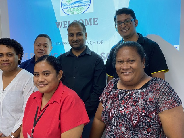
Board Members

Fiji COE
In October the Fiji WFUMB CoE ran a very successful head & neck course.
Held at the Suva campus of the Fiji National University, the two day course was led by New Zealand sonographer Jo McCann and assisted by CoE regional director Sue Westerway from Sydney. The 27 participants attended from not only Fiji but also the surrounding Pacific nations of Tonga, Tavalu and Vanuatu.
Although quite advantageous in otolaryngology, ultrasound is rarely used for the head and neck region in the pacific, partly because of the structural complexity of the anatomy and the lack of education and training. The head and neck course was exceptionally facilitated and contributed significantly to upskilling ultrasound practitioners to promote the use of ultrasound. The course introduced participants to the techniques, protocols, and guidelines of head and neck imaging while also highlighting the common pathologies, their appearances, differentials, and reporting requirements of surgeons. The COE would be interested in running more head and neck ultrasound training over a longer duration for advanced ultrasound users.
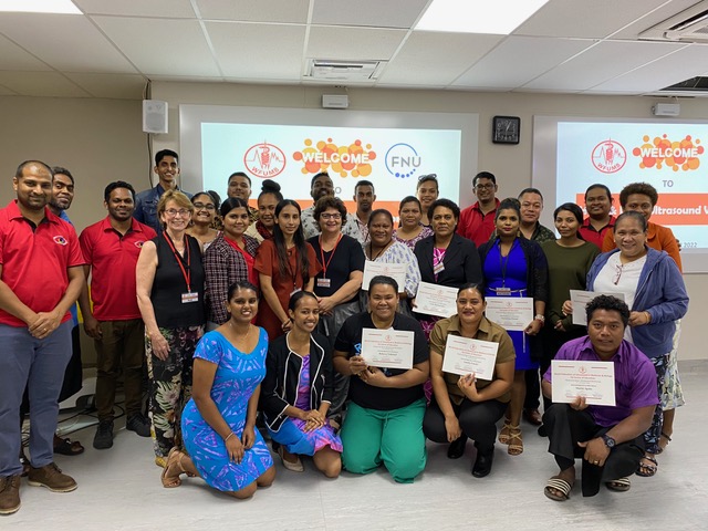
The participants with their certificates of attendance

2h Project in Cambodia
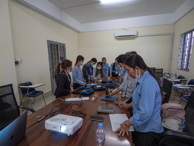
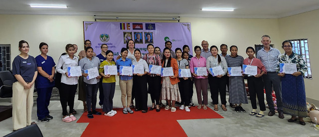
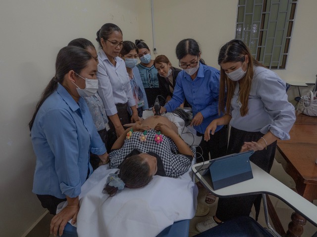
WFUMB were delighted to donate a Butterfly and LeSono device on the 2h Project
- Our main effort is to train a small cohort of midwives to use Butterfly ultrasound devices to take back to their health (delivery) centres. This has a clear purpose. Ensure women with pregnancies at risk of delivery complications, deliver in the safety of the local hospital. The midwives are flying after two days. We are thrilled so with their progress. They completed their theory and on-site training today. Angel is heading out to a health centre tomorrow to transition one set of midwives to their environment.
- Provide highly skilled scanning for the surgeon who has come with the project to ensure he is operating accurately and appropriately. (This has barely been needed)
- Train any local doctors in the use of ultrasound, There are no sonographers in Pailin so this has been uneven. The standard of scanning is very poor. There is no CT or MR so they are very dependent on ultrasound. Unfortunately there are strictly no photos around the hospital. Suffice to say, the patient experience is very poor (more on this on return.). There were a some moments today where we made a significant difference to a patient's outcome. The days have been book-ended with formal dinners, receptions and a breakfast. We haven't stopped.
Monash Health

ASUM News Report
ASUM Awards of Excellence 2022
The Australasian Society for Ultrasound in Medicine congratulates our 2022 Awards of Excellence winners. In addition, we acknowledge all those who were nominated as they strive to deliver ultrasound excellence.
ASUM Drive: Digital Library
ASUM Drive, our digital on-demand library, delivers digital content in a modern and easily accessible way. Our members receive this complimentary, and the feedback has been overwhelmingly positive. We continue to build on this resource with over 500 presentations available for your learning pleasure. This resource is available for the wider community with an annual subscription or rent.
ASUM launched the parent-centred communication in obstetric ultrasound guidelines earlier this year. Healthcare professionals and their practices are encouraged to discuss and develop policies and protocols to provide a consistent and transparent approach, utilising the best practice framework to optimise parent communication.
Inspiring Sonographers – Australasian Sonographers Day
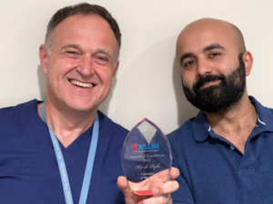 Australasian Sonographers Day, 27 November, celebrates inspiring sonographers such as 2022 Educator of the Year Rex de Ryke and rural heroes such as nominated Sonographer in Katherine, Chiharu Lupuleasa.
"Chiharu is now doing cardiac scans on top of her busy general sonography duties. Today she had an overbooked hospital inpatient and outpatient general list and was called to do an urgent echo on a sick patient.
This juggling happens in many places for sure, but they have the added pressure of isolation, in terms of geographical isolation and in person support."
 AJUM
AJUM
The Australasian Journal of Ultrasound in Medicine (AJUM) is a multi-disciplinary journal with an extensive international readership.
"When I started to learn sonography, I remember my training was informal, largely on patients, and very specific to the supervisor's skills and approaches. There was little standardisation and protocols, and much was 'borrowed’ from overseas departments where our first leaders and mentors had trained themselves."...
Read Professor Gillian Whalley's new free access article, 'Evolution in training in ultrasound' and much more in the just released AJUM November issue.
ASUM Outreach
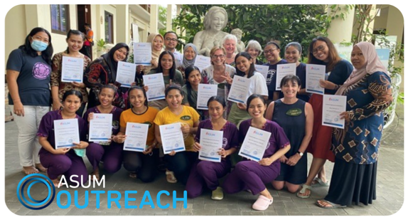
ASUM Outreach volunteer held a 4-day obstetric course that included lectures in Bahasa as well as pre-and post-course quizzes for 4 doctors and 14 nurses and midwives from Bali, Sumba, Papua & Lombok.
12 of the students were complete beginners but by the final day, all were able to complete an assessment for identifying anatomy and correctly measuring a fetal head, abdomen, and femur.
The doctors were also introduced to FAST to assist in their remote clinics.
Donated ultrasound equipment varied from old to hand-held, but by teaching clinic staff the basics of obstetric POCUS they can save lives.
Thanks to our dedicated trainers Sue Westerway, Debre Paoletti, Fiona Singer, and Sue Hewlett for continuing this important work.

EFSUMB Report
 EUROSON 2023
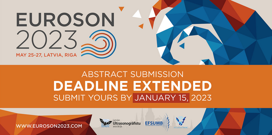
Pre-Congress School
The day before the congress, in addition we invite you to participate in the Pre-Congress School. There will be two parallel topics:
(I) Musculoskeletal ultrasound. Topics: Shoulder, Ankle and Foot with Live DEMOs by Prof. Carlo Martinoli and Faculty
(II) Contrast-enhanced ultrasound (CEUS). Topics: Liver and Non-Liver Applications and Case discussions by EFSUMB Faculty
You can get familiar with the preliminary program of the Pre-Congress School on the congress webpage: www.euroson2023.com/programme/ Please be aware that Pre-Congress School events are subject to the limited availability. Therefore, priority will be given to those participants who register earlier.
Registration to the Pre-Congress Schools is available here - www.euroson2023.com/register/
We are looking forward to seeing you in Riga!
EUROSON Schools

COE REPORT - Indonesia
Inauguration of Peru CoE
The Indonesian COE conducted two educational programmes in 2022.
Programme one was a Basic Thoracic Ultrasound Course which took place 20-21 September 2022 and was attended by 37 people. The second programme took place on 8-10 December 2022 and was The Internal Medicine Specialist Ultrasound Phase I, with 47 attendees. Both activities were well received and successful.
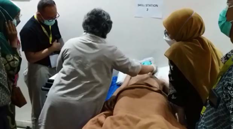
Basic Thoracic Ultrasound Course
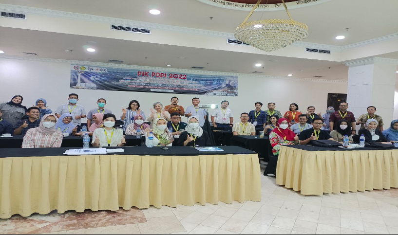
Attendees of Basic Thoracic Ultrasound Course
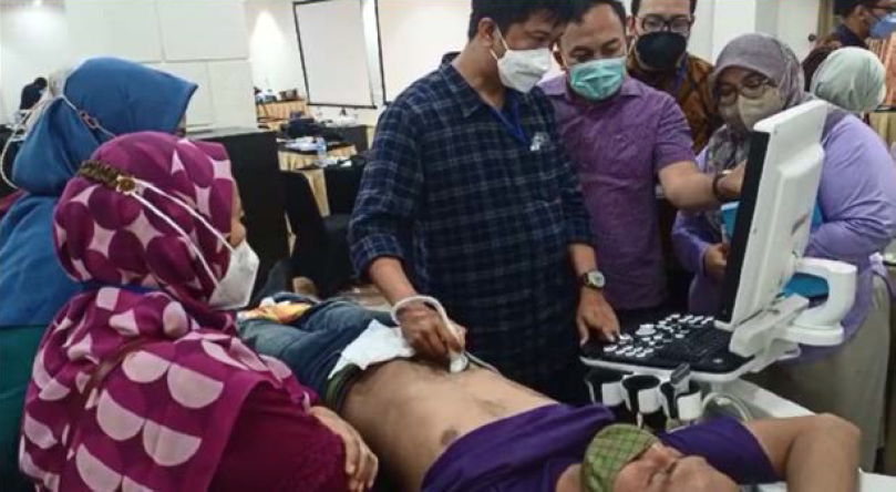
The Internal Medicine Specialist Ultrasound Phase I
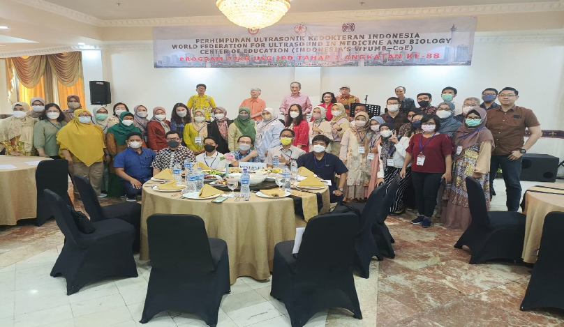
Attendees of The Internal Medicine Specialist Ultrasound Phase I
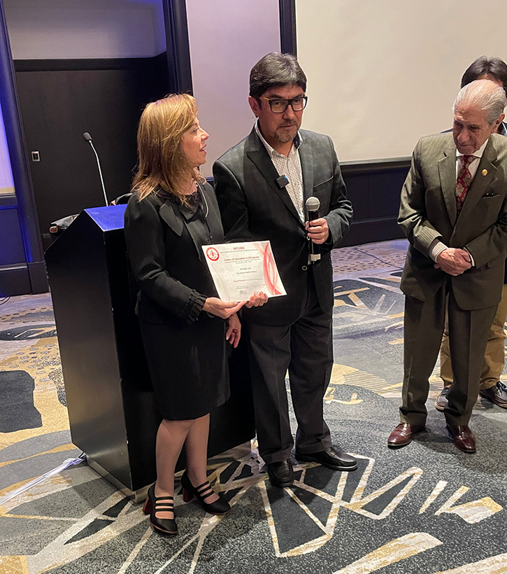
The official Inauguration of Peru CoE took place in October 2022.

Luminary Interview with David H Evans
Brief Biography
David H Evans is an Emeritus Professor of Medical Physics in the Department of Cardiovascular Sciences at the University of Leicester.
His principal research interests are related to Doppler ultrasound physics and signal processing, and applications of Doppler ultrasound in the cerebral circulation. He is the principal co-author of two textbooks on Doppler ultrasound physics, has authored over 200 peer reviewed papers, and lectured in 24 different countries around the world. He has served as President of the British Medical Ultrasound Society (1996-1998), President of European Federation of Societies for Ultrasound in Medicine and Biology (2005-2007), and Honorary Secretary of the World Federation for Ultrasound in Medicine and Biology (2009-2013). He is a Fellow of the Institute of Physics, and the Institute of Physics and Engineering in Medicine, an Honorary Fellow of the American Institute of Ultrasound in Medicine, and an Honorary Member of the British Medical Ultrasound Society.
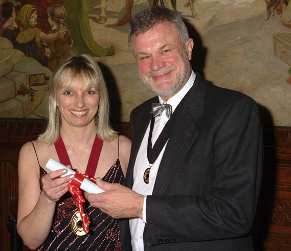
David receiving Honorary Membership of the British Medical Ultrasound Society from Jane Bates in 2004
How and when did you begin your ultrasound Career?
My career in ultrasound started just over 50 years ago, in September 1972, when I joined the Department of Medical Physics and Biomedical Engineering in Glasgow, Scotland. Even as a child I was fascinated by applications of technology in medicine and determined during my undergraduate physics course that I really wanted to apply my physics knowledge to medical problems. I didn’t specifically choose to go into ultrasound, but as an undergraduate I had spent an ‘Industrial Year’ working in the Acoustics Section of the UK National Physical Laboratory in Teddington, and so when I arrived in Glasgow as a new graduate it seemed only natural to set me to work with Norman McDicken on ultrasonics. After a fairly brief period in the central department in Glasgow I was sent to work with a group of vascular surgeons at the Glasgow Western Infirmary where I became particularly interested in Doppler ultrasound, something that has been the main focus of my work in ultrasound since that time.
Shortly after my move to the Western Infirmary Peter Bell, with whom I was then working, was appointed Professor of Surgery at the newly established Medical School in Leicester and I decided that I wanted to continue with the work I had started with Professor Bell, particularly as I thought that being involved at the new Medical School from the outset would present many new opportunities, and so moved to Leicester after just two years in Glasgow, and have continued to work in Leicester for most of the rest of my career.
What were your ultrasound career highlights?
If I were to be asked to name one highlight, it would be having the opportunity to work with so many talented and inspirational physicists, engineers, surgeons, neonatologists, and other clinicians, many of whom I now count amongst my close friends, and some of whom are sadly no longer with us. To be successful, a physicist working in medicine must forge a close working relationship with clinicians, and I have been extraordinarily lucky to have met many brilliant collaborators who have shaped my clinical interests.
Another highlight would be having the opportunity to travel widely, and lecture and teach in many different countries in many parts of the world, and to make many friends across the globe.
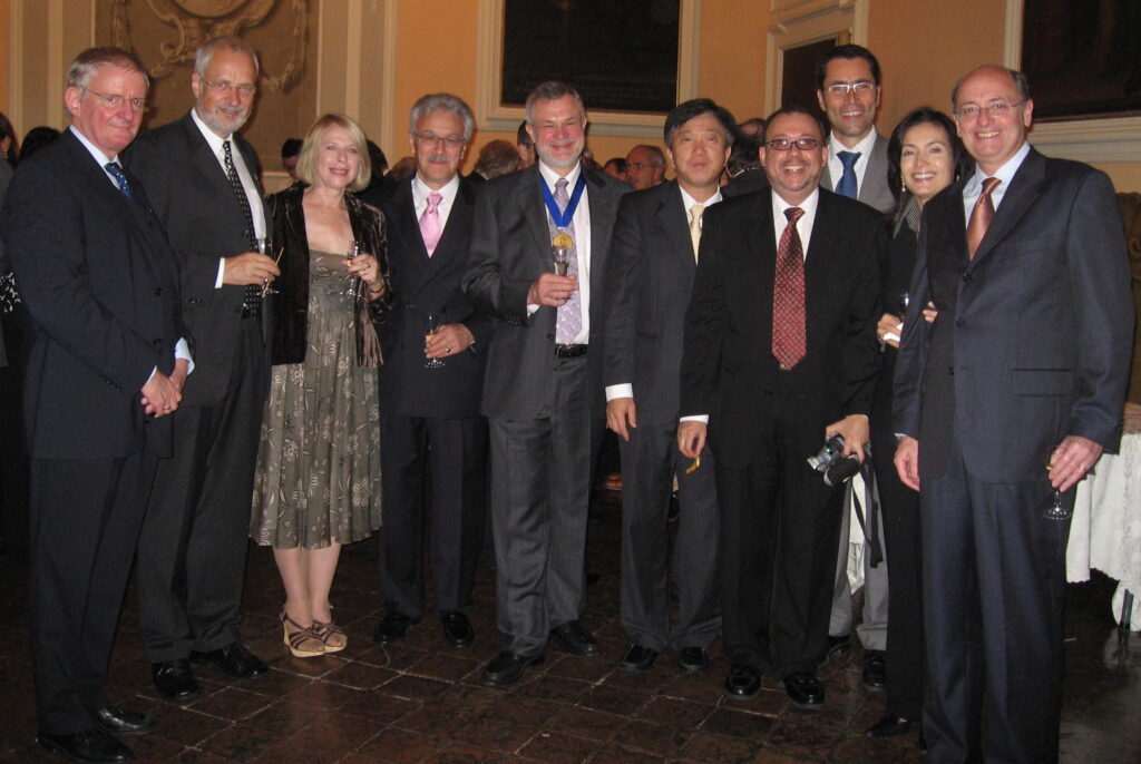
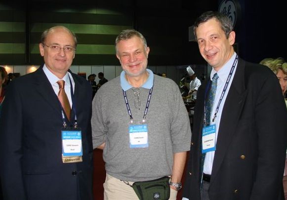
Some representatives at a Joint meeting of the EFSUMB and WFUMB Boards held in conjunction with Euroson 2006 in Bologna, Italy.
Other highlights have included working in Brazil (at the Federal University of Rio de Janeiro) for two years early on in my career, and of course being elected President of the British Medical Ultrasound Society, President of the European Federation of Societies for Ultrasound in Medicine and Biology, and Honorary Secretary of the World Federation for Ultrasound in Medicine and Biology.
What has been the most significant changes you have seen in ultrasound technology?
When I started working in ultrasound both imaging and Doppler were unbelievably primitive in comparison to today’s techniques. Individual B-scan images were formed over several seconds by exposing a polaroid film to the display of ultrasound echoes on a cathode ray oscilloscope as the ultrasound probe was moved over the surface of a patient’s body. Only after developing the film was it possible to see a full image, which would only display echoes from significant specular reflectors. The only way of imaging moving structures such as the heart was using M-mode displays. Doppler signals could only be interpreted in real-time either by ear, or by simple and unreliable envelope extraction techniques. Spectrum analysis of the signal was only possible using a ‘Kay Sona-Graph Sound Spectrograph’ which would take an extended period of time to analysis a 2.4 second sample of the Doppler signal and display its spectrum on thermal paper.
There have of course been many significant changes in ultrasound technology over the years, most of them have been incremental in nature, but some have been ‘game-changers’, and amongst these I would pick-out the introduction of grey-scale imaging by George Kossoff and his colleagues in Australia, the development of real-time scanning by a number of groups around the world, and the introduction of real-time colour flow imaging by Kasai, Namekawa, and their colleagues in Japan. The ability to display the spectral content of Doppler signals in real-time has also been a vital advancement in our ability to understand and interpret these signals. Ultrasound technology of course continues to improve and is now principally driven by improvements in transducer design and the ever-increasing power of computers.
What has been your most recent involvement with ultrasound?
Throughout my career I have been involved in many applications of ultrasound, notably in peripheral vascular disease, neonatology, the cerebral circulation, and embolus detection, and it is in these last two areas that I still have a particular interest. Today however my main contributions in these areas are in lecturing and writing of review articles and chapters.

Administrative News/Reminders
APISAMU
- A reminder to all affiliate organisations that nominations for committee members and EXB positions will be sent out in early 2023 for elections to be held in Riga, Latvia during EUROSON 2023 in 27 May.
- AIUM has affirmed that they will not be rejoining WFUMB at least for the next two years.
- WFUMB hopes to report favorably on meetings with SRU (Society of Radiologists in Ultrasound) who may consider forming a North American Ultrasound Federation which could include Canadian sonographers and ultrasound clinicians, leaving the way open for AIUM to return as part of this Federation. We will of course report further on this as our meetings progress.
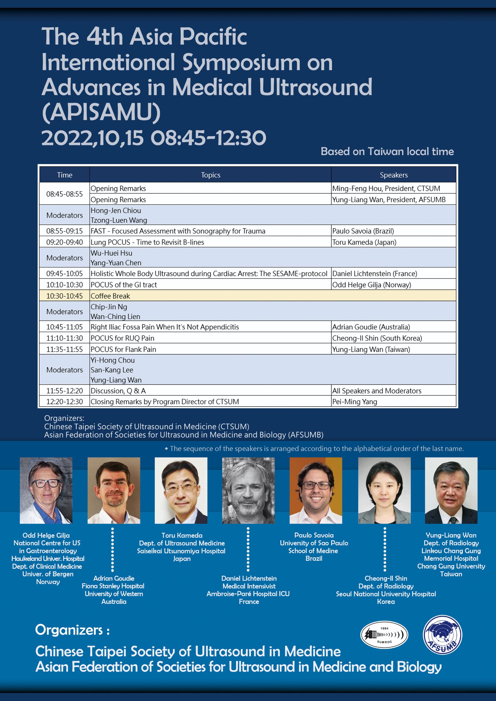
The 4th Asia Pacific International Symposium on Advances in Medical Ultrasound (APISAMU) was hosted by Taiwan Society of Ultrasound in Medicine and AFSUMB on October 15, 2022, with the theme of PoCUS. There were invited speakers from 7 countries.

In Memoriam: Dr. Beryl Benacerraf
 It is with great sadness that we learned of the passing of Dr. Beryl Benacerraf after a prolonged and courageous fight with esophageal cancer. The World Federation for Ultrasound in Medicine and Biology (WFUMB) offers our deepest condolences to her husband, Peter Libby, and her children, Brigitte and Oliver.
It is also with great sadness we remember our friend Dr Marvin Ziskin, WFUMB President 2003-2006, who passed away late this year. His obituary can be viewed here >. We ask you revisit the great Luminary interview with Dr Ziskin which we published in our March 2022 edition of ECHOES.

AFSUMB Report
Upcoming Events
The 15th onsite Congress of the AFSUMB 2022 was successfully held in Hyderabad, India from Nov. 11 to 13, 2022.
The Presidents of the congress were Dr. Sudheer Gokhale and Dr. Yung Liang Wan. The secretary was Dr. TLN Praveen. There was a total of 571 attendees including 475 domestic delegates and 96 international registrants. There were 106 faculty members. The academic schedule was spread over five parallel sessions. The LOC had arranged eight live demo workshops every day. There was a plenary session on the 12th which included five topics.
Despite the pandemic, the delegates or councilors from 11 out of 16 affiliations were able to attend the meeting.
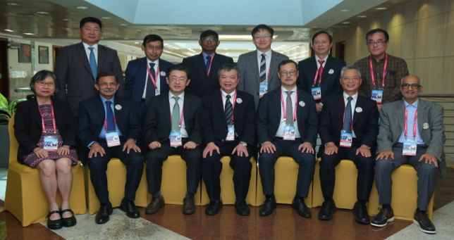
A group photo of the Officers and Administration councilors of the AFSUMB taken on Nov. 12, 2022, during the AFSUMB Congress at Hyderabad, India.
Left to right in the first row:
Chiou Li Ong (Singapore), Sudheer Gokhale (India), Jeong Yeon Cho (South Korea), Yung-Liang Wan (Taiwan), Iwaki Akiyama (Japan), Ching-Chang Hsieh (Taiwan), and Saqar Altai (Oman).
Left to right in the second row:
Erdenebileg Bavuujav (Mongolia), Kanu Bala (Bangladesh), TP Baskaran (Malaysia), Jae Young Lee (South Korea), Quan P.B. Nguyen (Vietnam), and Tuangsit Wataganara (Thailand).

