Caio Batalha Pereira, Simone Uezato Ota, Marcelo Schelini, Julia Diva Zavaris, Wagner Iared, Maria Cristina Chammas
- Centro de Aperfeiçoamento e Pesquisa em Ultrassonografia Prof. Dr. Giovanni Guido Cerri, DASA, São Paulo, Brazil.
Clinical History:
A 23-year-old womanfrom the countryside of São Paulo came to our clinicfora routine gynecologic examination. She had no known comorbidity.
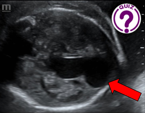
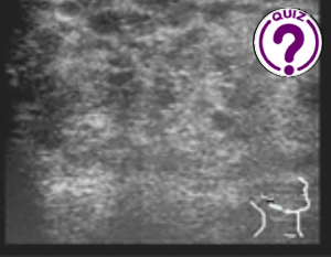
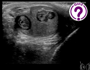
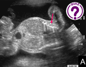
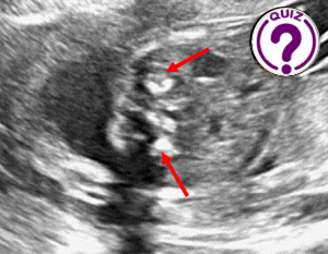
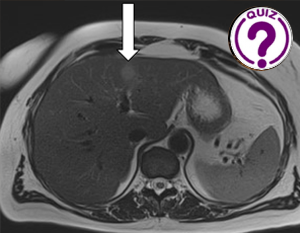
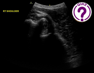
The figures represent:
Presence of ill-defined nodular formation, hyperechogenic, without Doppler flow, in the right ovary, measuring approximately 0.8 x 0.7 x 0.6 cm. Volume: 0.3 cm³.
Quiz-summary
0 of 1 questions completed
Questions:
- 1
Information
View the April Case below, answer the question and then click check >
You have already completed the quiz before. Hence you can not start it again.
Quiz is loading...
You must sign in or sign up to start the quiz.
You have to finish following quiz, to start this quiz:
Results
0 of 1 questions answered correctly
Your time:
Time has elapsed
You have reached 0 of 0 points, (0)
Categories
- Not categorized 0%
- 1
- Answered
- Review
-
Question 1 of 1
1. Question
Question: Which diagnosis is most likely for the case?
Correct
CORRECT ANSWER EXPLAINED BELOW Correct answer is: Dermoid cyst
Dermoid cysts account for 15% of all ovarian neoplasms and predominate in young women, being the most common ovarian neoplasm in patients younger than 20 years. Ultrasound is the preferred imaging modality. Typically, an ovarian dermoid is seen as a unilocular cystic adnexal mass with some mural components. The spectrum of sonographic features includes: a diffusely or partially echogenic mass with posterior sound attenuation owing to sebaceous material and hair within the cyst cavity; an echogenic interface at the edge of the mass that obscures deep structures (the tip of the iceberg sign); mural hyperechoic or Rokitansky nodule (dermoid plug); echogenic shadowing calcific or dental (tooth) components; fluid-fluid levels; multiple thin echogenic bands caused by the hair in the cyst cavity; the dot-dash pattern (dermoid mesh); no internal vascularity on color Doppler; intracystic floating balls sign (uncommon, but characteristic).
Discussion
Among the alternatives, the diagnosis of dermoid cyst should be considered because ovarian serous or mucinous cystadenoma/cystadenocarcinoma is usually only a serious consideration if the typical features of dermoid cyst are absent. Carcinoma also tends to occur in an older age group than dermoid cysts. Ovarian dysgerminomas are rare ovarian tumors that occur predominantly in young women in the 2nd to 3rd decades. Approximately 10-20% of cases occur in pregnancy. On ultrasound, they may be seen as a septated ovarian mass with varying echotexture. Color Doppler ultrasound may show prominent flow signal within the fibrovascular septa. Fibromas occur at all ages but are most frequently seen in middle-aged women, and on ultrasound manifest as solid, hypoechoic masses with ultrasound beam attenuation. As such, they may appear similar to a pedunculated subserosal uterine fibroid. However, the sonographic appearance can be variable, and some tumors can have cystic components, although rarely.
Take home message
Knowing the ultrasound aspects of dermoid cysts is important because they are benign lesions that grow slowly (1-2 mm per year) and, therefore may not need surgery. The most common complication is ovarian torsion, which occurs in 3-16% of dermoid cysts. Many recommend annual follow-up for lesions <7 cm to monitor growth. Tumors larger than 7 cm should be resected.
References
- Outwater EK, Siegelman ES, Hunt JL. Ovarian teratomas: tumor types and imaging characteristics. Radiographics. 21 (2): 475-90
- Adusumilli S, Hussain HK, Caoili EM et-al. MRI of sonographically indeterminate adnexal masses. AJR Am J Roentgenol. 2006;187 (3): 732-40.
- Gürel H, Gürel SA. Ovarian cystic teratoma with a pathognomonic appearance of multiple floating balls: a case report and investigation of common characteristics of the cases in the literature. Fertility and sterility. 90 (5): 2008.e17-9.
- Acién P, Ruiz-Maciá E, Acién M, Martín-Estefanía C. Mature ovarian teratoma-associated limbic encephalitis. Journal of obstetrics and gynaecology : the journal of the Institute of Obstetrics and Gynaecology. 35 (3): 317-9.
- Patel MD, Feldstein VA, Lipson SD et-al. Cystic teratomas of the ovary: diagnostic value of sonography. AJR Am J Roentgenol. 1998;171 (4): 1061-5
- Park SB, Kim JK, Kim KR et-al. Imaging findings of complications and unusual manifestations of ovarian teratomas. Radiographics. 28 (4): 969-83.
- Moomjian LN, Clayton RD, Carucci LR. A Spectrum of Entities That May Mimic Abdominopelvic Abscesses Requiring Image-guided Drainage. (2018) Radiographics : a review publication of the Radiological Society of North America, Inc. 38 (4): 1264-1281.
Incorrect
CORRECT ANSWER EXPLAINED BELOW Correct answer is: Dermoid cyst
Dermoid cysts account for 15% of all ovarian neoplasms and predominate in young women, being the most common ovarian neoplasm in patients younger than 20 years. Ultrasound is the preferred imaging modality. Typically, an ovarian dermoid is seen as a unilocular cystic adnexal mass with some mural components. The spectrum of sonographic features includes: a diffusely or partially echogenic mass with posterior sound attenuation owing to sebaceous material and hair within the cyst cavity; an echogenic interface at the edge of the mass that obscures deep structures (the tip of the iceberg sign); mural hyperechoic or Rokitansky nodule (dermoid plug); echogenic shadowing calcific or dental (tooth) components; fluid-fluid levels; multiple thin echogenic bands caused by the hair in the cyst cavity; the dot-dash pattern (dermoid mesh); no internal vascularity on color Doppler; intracystic floating balls sign (uncommon, but characteristic).
Discussion
Among the alternatives, the diagnosis of dermoid cyst should be considered because ovarian serous or mucinous cystadenoma/cystadenocarcinoma is usually only a serious consideration if the typical features of dermoid cyst are absent. Carcinoma also tends to occur in an older age group than dermoid cysts. Ovarian dysgerminomas are rare ovarian tumors that occur predominantly in young women in the 2nd to 3rd decades. Approximately 10-20% of cases occur in pregnancy. On ultrasound, they may be seen as a septated ovarian mass with varying echotexture. Color Doppler ultrasound may show prominent flow signal within the fibrovascular septa. Fibromas occur at all ages but are most frequently seen in middle-aged women, and on ultrasound manifest as solid, hypoechoic masses with ultrasound beam attenuation. As such, they may appear similar to a pedunculated subserosal uterine fibroid. However, the sonographic appearance can be variable, and some tumors can have cystic components, although rarely.
Take home message
Knowing the ultrasound aspects of dermoid cysts is important because they are benign lesions that grow slowly (1-2 mm per year) and, therefore may not need surgery. The most common complication is ovarian torsion, which occurs in 3-16% of dermoid cysts. Many recommend annual follow-up for lesions <7 cm to monitor growth. Tumors larger than 7 cm should be resected.
References
- Outwater EK, Siegelman ES, Hunt JL. Ovarian teratomas: tumor types and imaging characteristics. Radiographics. 21 (2): 475-90
- Adusumilli S, Hussain HK, Caoili EM et-al. MRI of sonographically indeterminate adnexal masses. AJR Am J Roentgenol. 2006;187 (3): 732-40.
- Gürel H, Gürel SA. Ovarian cystic teratoma with a pathognomonic appearance of multiple floating balls: a case report and investigation of common characteristics of the cases in the literature. Fertility and sterility. 90 (5): 2008.e17-9.
- Acién P, Ruiz-Maciá E, Acién M, Martín-Estefanía C. Mature ovarian teratoma-associated limbic encephalitis. Journal of obstetrics and gynaecology : the journal of the Institute of Obstetrics and Gynaecology. 35 (3): 317-9.
- Patel MD, Feldstein VA, Lipson SD et-al. Cystic teratomas of the ovary: diagnostic value of sonography. AJR Am J Roentgenol. 1998;171 (4): 1061-5
- Park SB, Kim JK, Kim KR et-al. Imaging findings of complications and unusual manifestations of ovarian teratomas. Radiographics. 28 (4): 969-83.
- Moomjian LN, Clayton RD, Carucci LR. A Spectrum of Entities That May Mimic Abdominopelvic Abscesses Requiring Image-guided Drainage. (2018) Radiographics : a review publication of the Radiological Society of North America, Inc. 38 (4): 1264-1281.