
Presentations from Webinar: Carotid Doppler: IMT, Stenosis and Cardiovascular Disease Impact
February 6, 2020test
February 24, 2020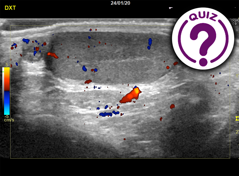
Scrotal pain
Jonathan Cohen, M. D.
Department of Radiology, Rigshospitalet – Copenhagen University Hospital, Denmark
Quiz-summary
0 of 1 questions completed
Questions:
- 1
Information
View the February Case below, answer the question and then click check >
You have already completed the quiz before. Hence you can not start it again.
Quiz is loading...
You must sign in or sign up to start the quiz.
You have to finish following quiz, to start this quiz:
Results
0 of 1 questions answered correctly
Your time:
Time has elapsed
You have reached 0 of 0 points, (0)
Categories
- Not categorized 0%
- 1
- Answered
- Review
-
Question 1 of 1
1. Question
Scrotal pain
Jonathan Cohen, M. D.
Department of Radiology, Rigshospitalet – Copenhagen University Hospital, DenmarkClinical History:
A 10-year-old boy was admitted to our hospital after 1.5 days of increasing right scrotal pain. The pain was present at all times, but the patient had been able to sleep. The second day the pain was intolerable, and the patient was sent home from school. He had never experienced similar pain and was generally in good health. He had neither urinary symptoms nor abdominal symptoms and no fever. Physical examination revealed pain on palpation at the level of the epididymis, with no scrotal swelling or redness and no signs of hernia or abdominal pain. A urinary dipstick test was negative.
Ultrasound Images:
The patient was examined with a high frequency linear transducer in the supine position including intermittent Valsalva maneuver. The attached images are from the examination. (Click on the images to enlarge)
-
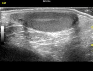
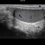
- Figure 1: Right scrotum, sagittal view.
-
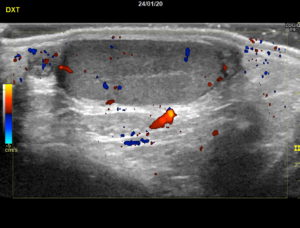
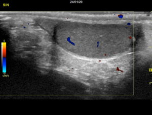
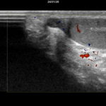
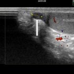
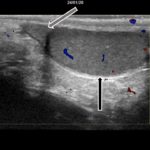
- Figure 2: Right scrotum, sagittal view, with colour Doppler. Slightly lateral to image 1.
-
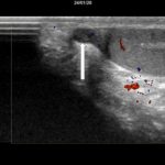
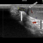
- Figure 3: Right scrotum, sagittal view, with colour Doppler.
-
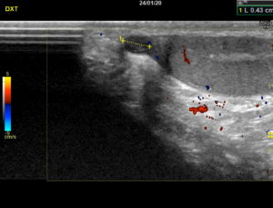
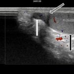
- Figure 4: Same as figure 3, with measurement.
-
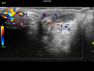
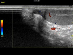
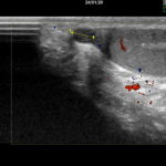
- Figure 5: Right scrotum, axial view, with colour Doppler
-
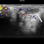
- Figure 6: Left side of the scrotum, sagital view.
Question: Which diagnosis is most likely?
Correct
CORRECT ANSWER EXPLAINED BELOW Ultrasound description
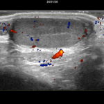
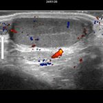
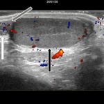
Ultrasound revealed a 4 mm, well circumscribed, slightly hypoechoic lesion in the right scrotum. It had a hyperechoic center and no flow on colour Doppler. The lesion did not change in size or location during Valsalva. The testis, epididymis and spermatic cord appeared normal.
Image gallery below indicates the original images without indicators, one with indicators indicating the lesion and one indicating both the lesion and adjacent anatomical structures. (click on images to enlarge)
-
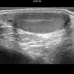
- Figure 1: Sagittal view without colour Doppler: Normal appearance of the testis and epididymis.
-
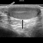
- Figure 1a: Black arrow: testis. Grey arrow: epididymis.
-
- Figure 2: Sagittal view. Probe slightly lateral to figure 1. Normal appearing testis and epididymis. Behind the epididymis, a slightly hypoechoic lesion with hyperechoic center is noted. There is no enhancement behind the lesion. Doppler flow is normal in the testis and epididymis. There is no free fluid in the scrotum.
-
- Figure 2a: White arrow: the lesion.
-
- Figure 2b: Black arrow: testis. White arrow: the lesion. Grey arrow: epididymis.
-
- Figure 3: Sagittal view. The lesion is seen on top of the epididymis. There is no flow on colour Doppler.
-
- Figure 3a: White arrow: the lesion.
-
- Figure 3b: Black arrow: testis. White arrow: the lesion. Grey arrow: epididymis.
-
- Figure 4: Same as 3 with measurement.
-
- Figure 4a: white arrow indicating the lesion with measurement.
-
- Figure 4b: Black arrow: testis. White arrow: the lesion. Grey arrow: epididymis.
-
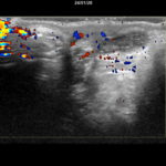
- Figure 5: Axial view. Close up of the hypoechoic lesion in the right side of scrotum next to the epididmymis at the upper pole of the right testis. There is no flow on colour Doppler.
-
- Figure 5a: White arrow: the lesion.
-
- Figure 6: Sagittal view: Normal appearance of the left testis and epididymis.
-
- Figure 6a: Black arrow: testis. Grey arrow: epididymis.
Discussion
The appendix epididymis is, along with the appendix testis, thought to be a vestigial remnant without vital function. While the appendix epididymis is a small, stalked structure attached to the head of the epididymis, the appendix testis is a fixed, globular structure located at the cranial pole of the testis[1,2]. The appendages may appear unassuming, but are clinically significant, as up to 60% of cases of acute scrotum in children have been reported to arise from torsion of the appendages [3,4].
The most common ultrasound findings in the case of a torsed appendix are the presence of an enlarged, hetero- or homogenous avascular mass with hyperemia and edema of the surrounding structures [2,3]. The condition most often requires only symptomatic treatment [3].
Discerning a relatively benign torsion of the appendages from the surgical emergency of testicular torsion by clinical examination is difficult, as symptoms often overlap. Ultrasound is considered to be both sensitive and specific in this regard, and findings suggestive of testicular torsion include absent or reduced intra-testicular blood flow compared with the asymptomatic side, twisting of the spermatic cord and abnormal location and appearance of the epididymal head [5].
Teaching points
Torsion of the epididymal or testicular appendix is one of the most common finding in the acute scrotum in children. Ultrasound examination with colour Doppler is an important tool in ruling out the more severe diagnosis of testicular torsion.
References
- Sahni D, Jit I, Joshi K, Sanjeev. Incidence and structure of the appendices of the testis and epididymis. J Anat. 1996 Oct;189 ( Pt 2):341–8.
- Sellars MEK, Sidhu PS. Ultrasound appearances of the testicular appendages: Pictorial review. Vol. 13, European Radiology. 2003. p. 127–35.
- Lev M, Ramon J, Mor Y, Jacobson JM, Soudack M. Sonographic appearances of torsion of the appendix testis and appendix epididymis in children. J Clin Ultrasound. 2015 Oct 1;43(8):485–9.
- Kalfa N, Veyrac C, Lopez M, Lopez C, Maurel A, Kaselas C, et al. Multicenter assessment of ultrasound of the spermatic cord in children with acute scrotum. J Urol. 2007 Jan;177(1):297–301; discussion 301.
- Alkhori NA, Barth RA. Pediatric scrotal ultrasound: review and update. Vol. 47, Pediatric Radiology. Springer Verlag; 2017. p. 1125–33.
Incorrect
CORRECT ANSWER EXPLAINED BELOW Ultrasound description
Ultrasound revealed a 4 mm, well circumscribed, slightly hypoechoic lesion in the right scrotum. It had a hyperechoic center and no flow on colour Doppler. The lesion did not change in size or location during Valsalva. The testis, epididymis and spermatic cord appeared normal.
Image gallery below indicates the original images without indicators, one with indicators indicating the lesion and one indicating both the lesion and adjacent anatomical structures. (click on images to enlarge)
-
- Figure 1: Sagittal view without colour Doppler: Normal appearance of the testis and epididymis.
-
- Figure 1a: Black arrow: testis. Grey arrow: epididymis.
-
- Figure 2: Sagittal view. Probe slightly lateral to figure 1. Normal appearing testis and epididymis. Behind the epididymis, a slightly hypoechoic lesion with hyperechoic center is noted. There is no enhancement behind the lesion. Doppler flow is normal in the testis and epididymis. There is no free fluid in the scrotum.
-
- Figure 2a: White arrow: the lesion.
-
- Figure 2b: Black arrow: testis. White arrow: the lesion. Grey arrow: epididymis.
-
- Figure 3: Sagittal view. The lesion is seen on top of the epididymis. There is no flow on colour Doppler.
-
- Figure 3a: White arrow: the lesion.
-
- Figure 3b: Black arrow: testis. White arrow: the lesion. Grey arrow: epididymis.
-
- Figure 4: Same as 3 with measurement.
-
- Figure 4a: white arrow indicating the lesion with measurement.
-
- Figure 4b: Black arrow: testis. White arrow: the lesion. Grey arrow: epididymis.
-
- Figure 5: Axial view. Close up of the hypoechoic lesion in the right side of scrotum next to the epididmymis at the upper pole of the right testis. There is no flow on colour Doppler.
-
- Figure 5a: White arrow: the lesion.
-
- Figure 6: Sagittal view: Normal appearance of the left testis and epididymis.
-
- Figure 6a: Black arrow: testis. Grey arrow: epididymis.
Discussion
The appendix epididymis is, along with the appendix testis, thought to be a vestigial remnant without vital function. While the appendix epididymis is a small, stalked structure attached to the head of the epididymis, the appendix testis is a fixed, globular structure located at the cranial pole of the testis[1,2]. The appendages may appear unassuming, but are clinically significant, as up to 60% of cases of acute scrotum in children have been reported to arise from torsion of the appendages [3,4].
The most common ultrasound findings in the case of a torsed appendix are the presence of an enlarged, hetero- or homogenous avascular mass with hyperemia and edema of the surrounding structures [2,3]. The condition most often requires only symptomatic treatment [3].
Discerning a relatively benign torsion of the appendages from the surgical emergency of testicular torsion by clinical examination is difficult, as symptoms often overlap. Ultrasound is considered to be both sensitive and specific in this regard, and findings suggestive of testicular torsion include absent or reduced intra-testicular blood flow compared with the asymptomatic side, twisting of the spermatic cord and abnormal location and appearance of the epididymal head [5].
Teaching points
Torsion of the epididymal or testicular appendix is one of the most common finding in the acute scrotum in children. Ultrasound examination with colour Doppler is an important tool in ruling out the more severe diagnosis of testicular torsion.
References
- Sahni D, Jit I, Joshi K, Sanjeev. Incidence and structure of the appendices of the testis and epididymis. J Anat. 1996 Oct;189 ( Pt 2):341–8.
- Sellars MEK, Sidhu PS. Ultrasound appearances of the testicular appendages: Pictorial review. Vol. 13, European Radiology. 2003. p. 127–35.
- Lev M, Ramon J, Mor Y, Jacobson JM, Soudack M. Sonographic appearances of torsion of the appendix testis and appendix epididymis in children. J Clin Ultrasound. 2015 Oct 1;43(8):485–9.
- Kalfa N, Veyrac C, Lopez M, Lopez C, Maurel A, Kaselas C, et al. Multicenter assessment of ultrasound of the spermatic cord in children with acute scrotum. J Urol. 2007 Jan;177(1):297–301; discussion 301.
- Alkhori NA, Barth RA. Pediatric scrotal ultrasound: review and update. Vol. 47, Pediatric Radiology. Springer Verlag; 2017. p. 1125–33.
-