Case of the Month February 2021- Medial knee pain in runner
February 2, 2021
Ultrasound the Best #20: Superficial and Skin Lumps/Nodes
March 8, 2021Caio Batalha Pereira, Simone Uezato Ota, Marcelo Schelini, Wagner Iared, Maria Cristina Chammas
- Centro de Aperfeiçoamento e Pesquisa em Ultrassonografia Prof. Dr. Giovanni Guido Cerri, DASA, São Paulo, Brazil.
Clinical History:
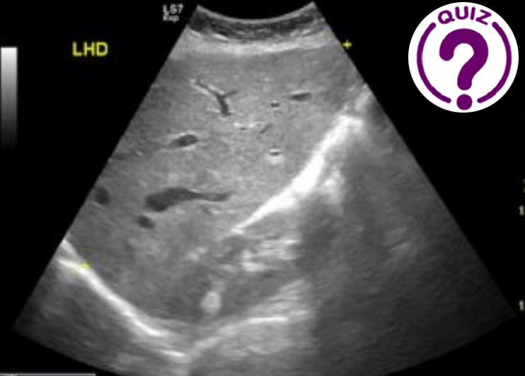
A 67-year-old woman from the countryside of São Paulo came to our clinic with complaints of mild right upper quadrant abdominal pain and slightly elevated serum liver enzymes. She had heart failure as comorbidity.
Quiz-summary
0 of 1 questions completed
Questions:
- 1
Information
View the March Case below, answer the question and then click check >
You have already completed the quiz before. Hence you can not start it again.
Quiz is loading...
You must sign in or sign up to start the quiz.
You have to finish following quiz, to start this quiz:
Results
0 of 1 questions answered correctly
Your time:
Time has elapsed
You have reached 0 of 0 points, (0)
Categories
- Not categorized 0%
- 1
- Answered
- Review
-
Question 1 of 1
1. Question
Question: Which diagnosis is most likely for the case?
Correct
CORRECT ANSWER EXPLAINED BELOW Correct answer is: Passive hepatic congestion.
Discussion
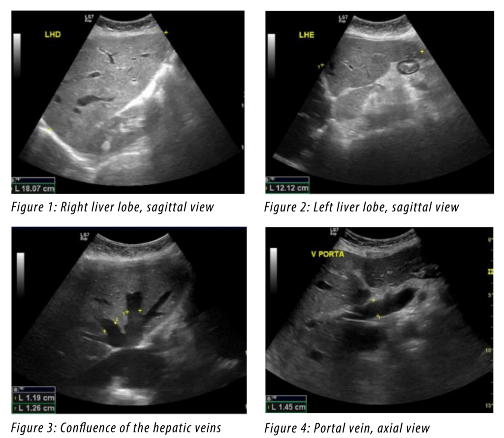
The figures show a slightly enlarged liver with regular contours and blunt edges. The liver echogenecity is homogeneous. There are no focal lesions. Enlarged hepatic veins, measuring 1.3 to 2 cm near the IVC. Enlarged portal vein with a diameter of 1.5 cm and without thrombi.
Passive hepatic congestion, also known as congested liver in cardiac disease, describes the stasis of blood in the hepatic parenchyma, due to impaired hepatic venous drainage, which leads to the dilation of the central hepatic veins and hepatomegaly. Passive hepatic congestion is a well-studied result of acute or chronic right-sided heart failure.
The spectrum of sonographic features includes enlargement of the right hepatic lobe and the right hepatic vein measures about 9 mm (normal caliber < 6 mm), increases up to 13 mm with pericardial effusion, dilated IVC/hepatic veins, hepatomegaly, ± ascites.
Expected findings related to alternative answers
Among the alternatives, the diagnosis of congestive hepatic congestion should be considered by the patient history and the imaging findings. Budd-Chiari syndrome refers to the clinical picture that occurs when there is partial or complete obstruction of the hepatic veins. It is characterized on imaging by ascites, caudate hypertrophy, peripheral atrophy and prominent collateral veins. Hepatic veno-occlusive disease (VOD), also known as sinusoidal obstruction syndrome (SOS), is a condition arising from occlusion of the hepatic venules. Ultrasound is the imaging modality of choice which may show: hepatomegaly; portal vein abnormalities, such as portal vein dilatation, portal venous pulsatility, hepatofugal portal venous flow; gallbladder wall thickening (> 6-8 mm); and ascites. Acute hepatitis is a clinical diagnosis and the spectrum of sonographic features includes hepatomegaly (most sensitive sign) >15.5 cm at the midclavicular line; starry sky appearance; gallbladder wall thickening; periportal edema; accentuated brightness of the portal veinous walls; overall decreased echogenicity.
Take home message
Knowing the ultrasound aspects of hepatic congestion is important because it allows the clinician to direct the treatment to the basic cause – the heart failure.
References
- Zakim D, Boyer TD. Hepatology. Saunders. ISBN:0721648363.
- McNaughton DA, Abu-Yousef MM. Doppler US of the liver made simple. RadioGraphics 2011; 31(1): 161–188.
- Gore RM, Mathieu DG, White EM et-al. Passive hepatic congestion: cross-sectional imaging features. AJR Am J Roentgenol. 1994;162 (1): 71-5.
- Murphy FB, Steinberg HV, Shires GT et-al. The Budd-Chiari syndrome: a review. AJR Am J Roentgenol. 1986;147 (1): 9-15
- Bayraktar UD, Seren S, Bayraktar Y. Hepatic venous outflow obstruction: three similar syndromes. World J. Gastroenterol. 2007;13 (13): 1912-27
- Brancatelli G, Vilgrain V, Federle MP et-al. Budd-Chiari syndrome: spectrum of imaging findings. AJR Am J Roentgenol. 2007;188 (2): W168-76
Incorrect
CORRECT ANSWER EXPLAINED BELOW Correct answer is: Passive hepatic congestion.
Discussion
The figures show a slightly enlarged liver with regular contours and blunt edges. The liver echogenecity is homogeneous. There are no focal lesions. Enlarged hepatic veins, measuring 1.3 to 2 cm near the IVC. Enlarged portal vein with a diameter of 1.5 cm and without thrombi.
Passive hepatic congestion, also known as congested liver in cardiac disease, describes the stasis of blood in the hepatic parenchyma, due to impaired hepatic venous drainage, which leads to the dilation of the central hepatic veins and hepatomegaly. Passive hepatic congestion is a well-studied result of acute or chronic right-sided heart failure.
The spectrum of sonographic features includes enlargement of the right hepatic lobe and the right hepatic vein measures about 9 mm (normal caliber < 6 mm), increases up to 13 mm with pericardial effusion, dilated IVC/hepatic veins, hepatomegaly, ± ascites.
Expected findings related to alternative answers
Among the alternatives, the diagnosis of congestive hepatic congestion should be considered by the patient history and the imaging findings. Budd-Chiari syndrome refers to the clinical picture that occurs when there is partial or complete obstruction of the hepatic veins. It is characterized on imaging by ascites, caudate hypertrophy, peripheral atrophy and prominent collateral veins. Hepatic veno-occlusive disease (VOD), also known as sinusoidal obstruction syndrome (SOS), is a condition arising from occlusion of the hepatic venules. Ultrasound is the imaging modality of choice which may show: hepatomegaly; portal vein abnormalities, such as portal vein dilatation, portal venous pulsatility, hepatofugal portal venous flow; gallbladder wall thickening (> 6-8 mm); and ascites. Acute hepatitis is a clinical diagnosis and the spectrum of sonographic features includes hepatomegaly (most sensitive sign) >15.5 cm at the midclavicular line; starry sky appearance; gallbladder wall thickening; periportal edema; accentuated brightness of the portal veinous walls; overall decreased echogenicity.
Take home message
Knowing the ultrasound aspects of hepatic congestion is important because it allows the clinician to direct the treatment to the basic cause – the heart failure.
References
- Zakim D, Boyer TD. Hepatology. Saunders. ISBN:0721648363.
- McNaughton DA, Abu-Yousef MM. Doppler US of the liver made simple. RadioGraphics 2011; 31(1): 161–188.
- Gore RM, Mathieu DG, White EM et-al. Passive hepatic congestion: cross-sectional imaging features. AJR Am J Roentgenol. 1994;162 (1): 71-5.
- Murphy FB, Steinberg HV, Shires GT et-al. The Budd-Chiari syndrome: a review. AJR Am J Roentgenol. 1986;147 (1): 9-15
- Bayraktar UD, Seren S, Bayraktar Y. Hepatic venous outflow obstruction: three similar syndromes. World J. Gastroenterol. 2007;13 (13): 1912-27
- Brancatelli G, Vilgrain V, Federle MP et-al. Budd-Chiari syndrome: spectrum of imaging findings. AJR Am J Roentgenol. 2007;188 (2): W168-76


