- WFUMB Stands with EFSUMB: stop bloodshed in Ukraine - read our statement here >
Case of the Month February 2021- Medial knee pain in runner
WFUMB E-Book_ch8_v8.2
January 29, 2021
Case of the Month March 2021- A heart liver
March 1, 2021Matthew Gourlay
- Fowler Simmons Radiology. Adelaide, South Australia
Clinical History:
57 year old female runner. Recent increase in running load with increasing medial knee pain.
Quiz-summary
0 of 1 questions completed
Questions:
- 1
Information
View the February Case below, answer the question and then click check >
You have already completed the quiz before. Hence you can not start it again.
Quiz is loading...
You must sign in or sign up to start the quiz.
You have to finish following quiz, to start this quiz:
Results
0 of 1 questions answered correctly
Your time:
Time has elapsed
You have reached 0 of 0 points, (0)
Categories
- Not categorized 0%
- 1
- Answered
- Review
-
Question 1 of 1
1. Question
Question: What is the likely diagnosis?
Correct
CORRECT ANSWER EXPLAINED BELOW Correct answer is: Medial tibial plateau stress fracture.
Discussion
Stress fractures of the lower limb are not an uncommon finding in runners. This may be due to an increase in load or training errors. They may also occur due to decreased bone density of individuals without necessarily any increase in loading.
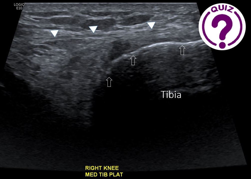
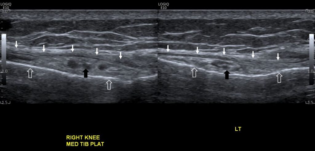
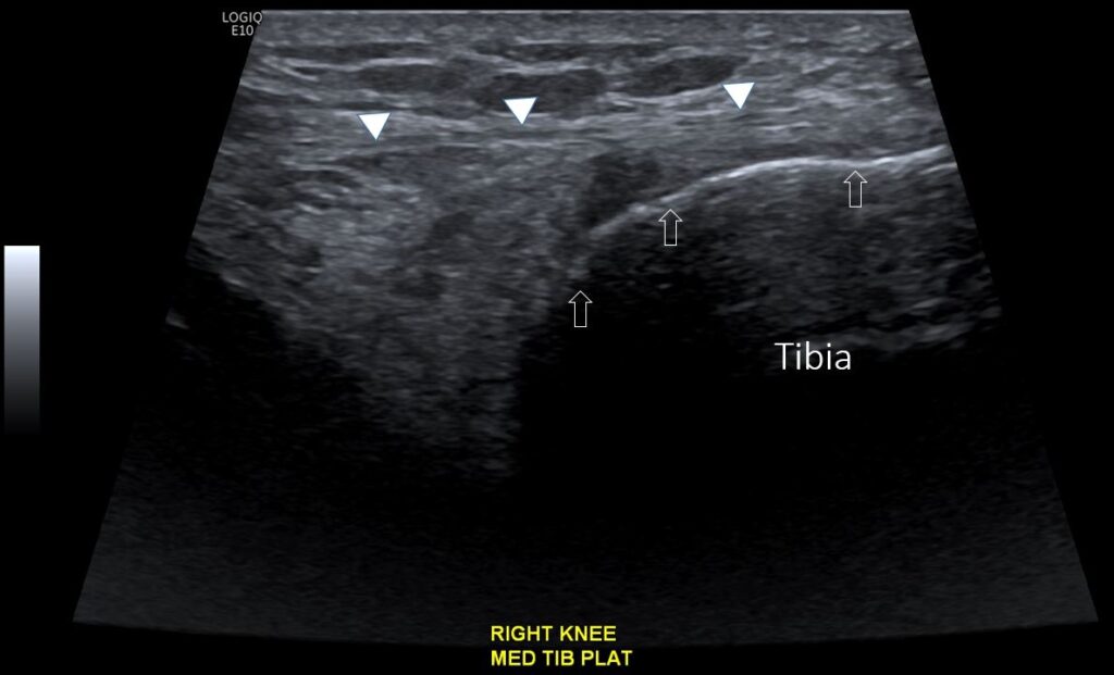
Our patient indicated an increase in loading along with tenderness over medial tibial plateau. On ultrasound assessment oedema of the fat layer overlying bone was noted along with periosteal haematoma which is shown as a hypoechoic line overlying the bone cortex (this is nicely shown in video 1 and Image 4). Swelling and oedema of the overlying fat can be noted on Image 1 between the medial collateral ligament and tibia. Clinical history and examination along with ultrasound findings are strongly indicative of stress fracture.
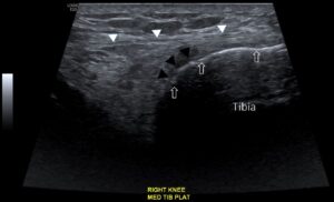
Image 4- Medial tibial plateau transverse view. Tibia (open white arrow), deep fascia (white arrow), periosteal haematoma (black arrow). Associated fat oedema can be noted overlying periosteal haematoma.
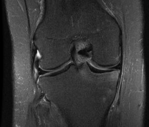
Image 3- PDFS MRI sequence showing bone marrow oedema of tibial plateau consistent with stress fracture.
Expected findings related to alternative answers
- A) Pes anserine bursitis-very rare to occur in isolation and swelling in this region can usually be attributed to one of the other 3 possible answers.
- B) Medial meniscal tear-clinically would present with pain and tenderness centred more over medial joint line rather than pes anserine region. Fluid and oedema uncommonly seen tracking down towards the pes anserine region due to a related meniscal cyst with or without rupture. Periosteal haematoma would not be present.
- D) Pes anserine bursitis may occur in patients with systemic arthropathy. This would result in localised swelling of tissues between medial collateral ligament (MCL) and pes anserine (unlike our current case which shows swelling under MCL).
Incorrect
CORRECT ANSWER EXPLAINED BELOW Correct answer is: Medial tibial plateau stress fracture.
Discussion
Stress fractures of the lower limb are not an uncommon finding in runners. This may be due to an increase in load or training errors. They may also occur due to decreased bone density of individuals without necessarily any increase in loading.
Our patient indicated an increase in loading along with tenderness over medial tibial plateau. On ultrasound assessment oedema of the fat layer overlying bone was noted along with periosteal haematoma which is shown as a hypoechoic line overlying the bone cortex (this is nicely shown in video 1 and Image 4). Swelling and oedema of the overlying fat can be noted on Image 1 between the medial collateral ligament and tibia. Clinical history and examination along with ultrasound findings are strongly indicative of stress fracture.

Image 4- Medial tibial plateau transverse view. Tibia (open white arrow), deep fascia (white arrow), periosteal haematoma (black arrow). Associated fat oedema can be noted overlying periosteal haematoma.

Image 3- PDFS MRI sequence showing bone marrow oedema of tibial plateau consistent with stress fracture.
Expected findings related to alternative answers
- A) Pes anserine bursitis-very rare to occur in isolation and swelling in this region can usually be attributed to one of the other 3 possible answers.
- B) Medial meniscal tear-clinically would present with pain and tenderness centred more over medial joint line rather than pes anserine region. Fluid and oedema uncommonly seen tracking down towards the pes anserine region due to a related meniscal cyst with or without rupture. Periosteal haematoma would not be present.
- D) Pes anserine bursitis may occur in patients with systemic arthropathy. This would result in localised swelling of tissues between medial collateral ligament (MCL) and pes anserine (unlike our current case which shows swelling under MCL).


