WFUMB Ultrasound Workshop Day: 28th May 2021
June 7, 2021
Student Education Committee MASUGiovanna Ferraioli
June 8, 2021Douglas Almeida de Oliveira Filho, MD
Thiago Adler Ralho Rodrigues dos Santos, MD, PhD
- Department of Radiology, University Hospital, Federal University of Mato Grosso do Sul (UFMS) Campo Grande –Brazil
Clinical History:
A 51-year-oldman complains about an increasing enlargement of the right testicle after a trauma 6 months ago. The physical examination showed a hardened painless right testicle, with volumetric increase, but no other signs or symptoms.
Quiz-summary
0 of 1 questions completed
Questions:
- 1
Information
View the June Case below, answer the question and then click check >
You have already completed the quiz before. Hence you can not start it again.
Quiz is loading...
You must sign in or sign up to start the quiz.
You have to finish following quiz, to start this quiz:
Results
0 of 1 questions answered correctly
Your time:
Time has elapsed
You have reached 0 of 0 points, (0)
Categories
- Not categorized 0%
- 1
- Answered
- Review
-
Question 1 of 1
1. Question
Question: Which diagnosis is most likely?
Correct
CORRECT ANSWER EXPLAINED BELOW Correct answer is: Testicular tumor
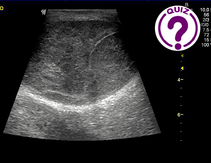
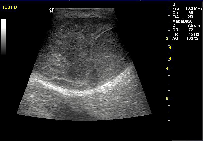
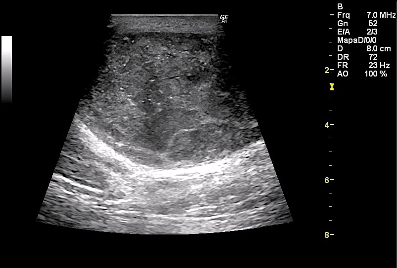
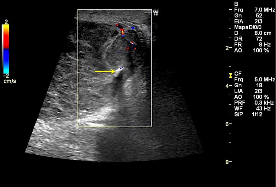
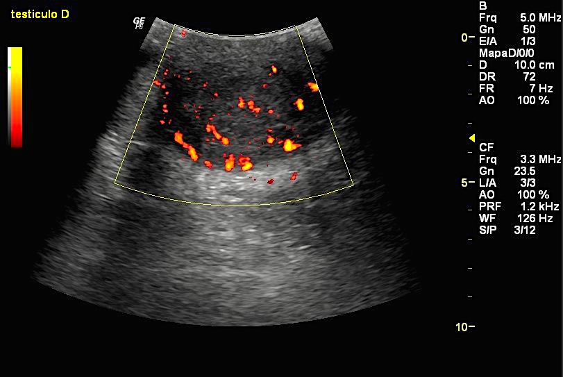
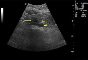
Ultrasound demonstrated an enlarged right testicle of128 ml (Fig. 1 and 2). The parenchyma was hypoechoic and heterogeneous, with small calcifications and regular contours (Fig. 3). Doppler imaging showed increased vascularization (Fig.4). Due to the suspicion of a testicular tumor in the scrotum, an abdominal ultrasound was also performed. This revealed a hypoechoic 4.6cm tumor with irregular contours in the right para-aortic region –just above the aortoiliac bifurcation –highly suspect of a metastatic lymph node (Fig.5).
Discussion
A testicular tumor should be suspected when an ultrasound shows a solid intratesticular mass with internal vascularization. It is essential to evaluate extra-testicular sites for lymph node metastases as disseminated metastases from testicular cancer may be seen here before the testiculartumor is identified(1). Especially, the retroperitoneal lymph nodes nearthe abdominal aorta should be examinedcarefully, as it is the main site for lymphatic spread(2). Infarction and hematoma are also relevant differential diagnoses when a mass is seen; however, no Doppler flow will be seen. In the case of infection, we would see an increase in Doppler flow in the affected testicle, but without the presence of a mass(3).
Teaching Points
The gold standard test for initial detection of testicular tumoris ultrasound, with a sensitivity close to 100% and specificity of 95-99%(3,4). Extra-testicular evaluation should be encouraged when malignancy is suspected.
Figure Legends
Figure 1 and 2 -Scrotal ultrasound performed with a linear transducer in the transversal plane (Fig. 1) and longitudinal plane (Fig. 2) demonstrated an enlarged right testicle with hypoechoic and diffuse heterogeneous echotexture and small hyperechoic focicorresponding tomicrocalcifications.
Figure 3 -Presence of a hyperechoic lesioninside the right testicle, suggestive of calcification (arrow).
Figure 4 -Scrotal ultrasound performed with a convex array transducer in the longitudinal plane demonstrated a diffuse increase in vascularization of the right testis on amplitude Doppler.
Figure 5 -Abdominal ultrasound performed with a convex array transducer, cross-section,showing the image of a right para-aortic lymph node, with loss of normal architecture, irregular contours (arrow) and the aorta (arrowhead).
References
- Moreno CC, Small WC, Camacho JC, et al. Testicular Tumors: What Radiologists Need to Know—Differential Diagnosis, Staging, and Management. RadioGraphics 2015; 35:400–415.
- Marko J, Wolfman DJ, Aubin AL, Sesterhenn IA. Testicular Seminoma and Its Mimics. RadioGraphics 2017;37:1085–1098.
- S. Gallego García, C. Santos Montón, I. Alonso Diego, et al. Testicular tumors: ultrasound diagnosis. European Society of Radiology. ECR 2019 / C-0935. 2019. DOI: 10.26044/ecr2019/C-0935
- Sumat K, Rumana M and Parveen Shah. Clinicopathological Characteristics of Testicular Tumors. Medwin Publishers. Clinical Pathology 2017, 1(1): 000106
Incorrect
CORRECT ANSWER EXPLAINED BELOW Correct answer is: Testicular tumor
Ultrasound demonstrated an enlarged right testicle of128 ml (Fig. 1 and 2). The parenchyma was hypoechoic and heterogeneous, with small calcifications and regular contours (Fig. 3). Doppler imaging showed increased vascularization (Fig.4). Due to the suspicion of a testicular tumor in the scrotum, an abdominal ultrasound was also performed. This revealed a hypoechoic 4.6cm tumor with irregular contours in the right para-aortic region –just above the aortoiliac bifurcation –highly suspect of a metastatic lymph node (Fig.5).
Discussion
A testicular tumor should be suspected when an ultrasound shows a solid intratesticular mass with internal vascularization. It is essential to evaluate extra-testicular sites for lymph node metastases as disseminated metastases from testicular cancer may be seen here before the testiculartumor is identified(1). Especially, the retroperitoneal lymph nodes nearthe abdominal aorta should be examinedcarefully, as it is the main site for lymphatic spread(2). Infarction and hematoma are also relevant differential diagnoses when a mass is seen; however, no Doppler flow will be seen. In the case of infection, we would see an increase in Doppler flow in the affected testicle, but without the presence of a mass(3).
Teaching Points
The gold standard test for initial detection of testicular tumoris ultrasound, with a sensitivity close to 100% and specificity of 95-99%(3,4). Extra-testicular evaluation should be encouraged when malignancy is suspected.
Figure Legends
Figure 1 and 2 -Scrotal ultrasound performed with a linear transducer in the transversal plane (Fig. 1) and longitudinal plane (Fig. 2) demonstrated an enlarged right testicle with hypoechoic and diffuse heterogeneous echotexture and small hyperechoic focicorresponding tomicrocalcifications.
Figure 3 -Presence of a hyperechoic lesioninside the right testicle, suggestive of calcification (arrow).
Figure 4 -Scrotal ultrasound performed with a convex array transducer in the longitudinal plane demonstrated a diffuse increase in vascularization of the right testis on amplitude Doppler.
Figure 5 -Abdominal ultrasound performed with a convex array transducer, cross-section,showing the image of a right para-aortic lymph node, with loss of normal architecture, irregular contours (arrow) and the aorta (arrowhead).
References
- Moreno CC, Small WC, Camacho JC, et al. Testicular Tumors: What Radiologists Need to Know—Differential Diagnosis, Staging, and Management. RadioGraphics 2015; 35:400–415.
- Marko J, Wolfman DJ, Aubin AL, Sesterhenn IA. Testicular Seminoma and Its Mimics. RadioGraphics 2017;37:1085–1098.
- S. Gallego García, C. Santos Montón, I. Alonso Diego, et al. Testicular tumors: ultrasound diagnosis. European Society of Radiology. ECR 2019 / C-0935. 2019. DOI: 10.26044/ecr2019/C-0935
- Sumat K, Rumana M and Parveen Shah. Clinicopathological Characteristics of Testicular Tumors. Medwin Publishers. Clinical Pathology 2017, 1(1): 000106

