WFUMB Stands with EFSUMB: stop bloodshed in Ukraine -
read our statement here >
MENU
MENU
About
ABOUT WFUMB
Constitutions & Bylaws
Mission Statement
Organogram
Leadership
5 Year Strategic Plan [2024]
Annual Report 2023
WFUMB Tax Returns
Contact WFUMB
WFUMB MEMBERSHIP
Affiliates
Partnerships
Benefits of WFUMB Membership
UMB Subscriptions
Honorary Members
MANAGEMENT
Administrative Councillors
Executive Bureau
Management Team
Standard Operating Procedures
PASSWORD PROTECTED
Executive Bureau Docs
Committees Docs
COMMITTEES
Education Committee
Collaboration Committee
Constitution Committee
Investment Advisory Committee
Publications Committee
Safety Committee
Congress Committee
Student Education
COE Task Force
E-learning Task Force
Outreach & COE's
CURRENT COE's
Albania
Bangladesh
Beijing, China
Zagreb, Croatia
Dominican Republic
Ethiopia
Fiji
CURRENT COE's
Indonesia
Kenya
Moldova
Mongolia
Paraguay
Peru
Philippines
CURRENT COE's
Romania
Sudan
Togo
Uganda
Venezuela
Vietnam
Zambia
OUTREACH
About COE'S and Locations
WFUMB Scholarship
COE Image of the Month
COE Evaluation Form
Education
WFUMB RESOURCES
WFUMB Lectures
WFUMB Congress 2023 Lectures
WFUMB Congress 2022 Lectures
Upcoming Webinars
Webinar Archive
Interactive Case of the Month
Ultrasound the Best Cases
Young Investigator Award
WFUMB Babyworks Scan Trainer Demonstration
PARTNER & AFFILIATE RESOURCES
Affiliate Resources
ICUS Web Site and Education
ISUOG Partnership Resources
WHO Courses
Exam Technique Videos
US knowledge Quiz
History
WFUMB HISTORY
History of WFUMB & Ultrasound
WFUMB History of Congresses
WFUMB PEOPLE
Presidents
Pioneers
Obituary
Calendar
Publications
JOURNALS/NEWSLETTERS
WFUMB Journal UMB
UMB Publications
WFUMB Ultrasound Open
Echoes
Federation Journals
BOOKS
Books
External Books
WFUMB Ultrasound Book
OTHER
Focal Liver Lesions
Incidental Findings
Ultrasound Atlas
How to Perform CEUS
Safety
WFUMB
Safety Statements
Safety Publications
OTHER
Federation Safety Statements
Other Safety Statements
Official Statements
Guidelines
Statements
Position Papers
COVID-19
✕
Ultrasound the Best Cases
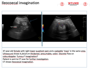
Ultrasound the Best #21: Ileocoecal Invagination
...
Read More
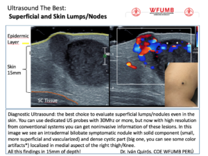
Ultrasound the Best #20: Superficial and Skin Lumps/Nodes
...
Read More
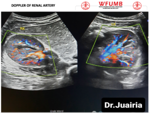
Ultrasound the Best #19: Doppler of Renal Artery
...
Read More
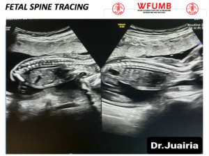
Ultrasound the Best #18: Fetal Spine Tracing
...
Read More
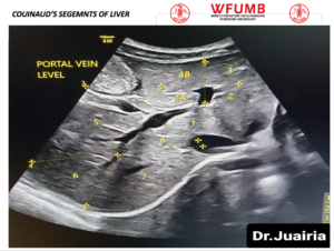
Ultrasound the Best #17: Segments of Liver
...
Read More
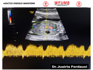
Ultrasound the Best #16: Ductus Venosus Waveform
...
Read More
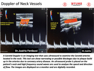
Ultrasound the Best #15: Doppler of Neck Vessels
...
Read More
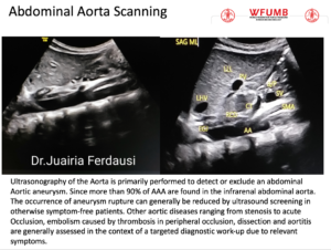
Ultrasound the Best #14: Abdominal Aorta Scanning
...
Read More
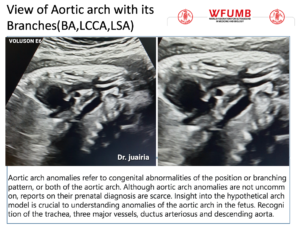
Ultrasound the Best #13: View of Aortic arch with its Branches (BA,LCCA,LSA)
...
Read More
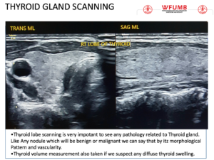
Ultrasound the Best #12: Thyroid gland scanning
...
Read More
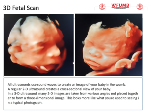
Ultrasound the Best #11: 3D Fetal Scan
...
Read More
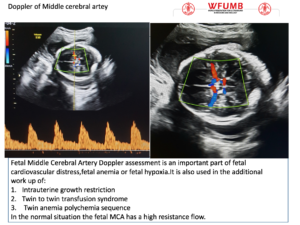
Ultrasound the Best #10: Doppler of the Fetal Middle of the Cerebral Artery
...
Read More
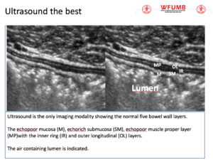
Ultrasound the Best #09: Ultrasound is the only imaging modality showing the normal five bowel wall layers.
...
Read More
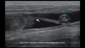
Ultrasound the Best #08: Appendix (in between markers) and mesappendix (arrow)
...
Read More
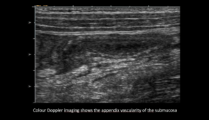
Ultrasound the Best #07: Colour Doppler imaging shows the appendix vascularity of the submucosa
...
Read More
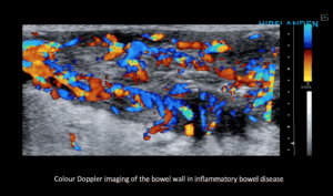
Ultrasound the Best #06: Colour Doppler imaging of the bowel wall in inflammatory bowel disease
...
Read More
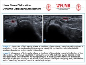
Ultrasound the Best #05: Ulnar Nerve Dislocation: Dynamic Ultrasound Assessment
...
Read More
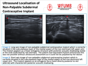
Ultrasound the Best #04: Ultrasound Localisation of Non-Palpable Subdermal Contraceptive Implant
...
Read More
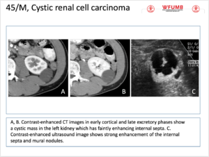
Ultrasound the Best #03: 45/M, Cystic renal cell carcinoma
...
Read More
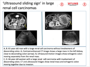
Ultrasound the Best #02: ‘Ultrasound sliding sign’ in large renal cell carcinomas
...
Read More
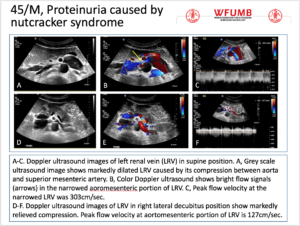
Ultrasound the Best #01: 45/M, Proteinuria caused by nutcracker syndrome
...
Read More
This website uses cookies to improve your experience. By using this website you agree to our
Data Protection Policy
.
Read more
Accept all
Translate »