
Webinar: Interventional Ultrasound (INVUS) – refresher and up-to-date information
March 6, 2020
WFUMB Position Statement: How to perform a safe ultrasound examination and clean equipment in the context of COVID-19
March 27, 2020
The Northern Lights CEUS Artifact
By Christian Pallson Nolsøe, MD, PhD
Zealand University Hospital, Køge, Department of Surgery
Clinical History:
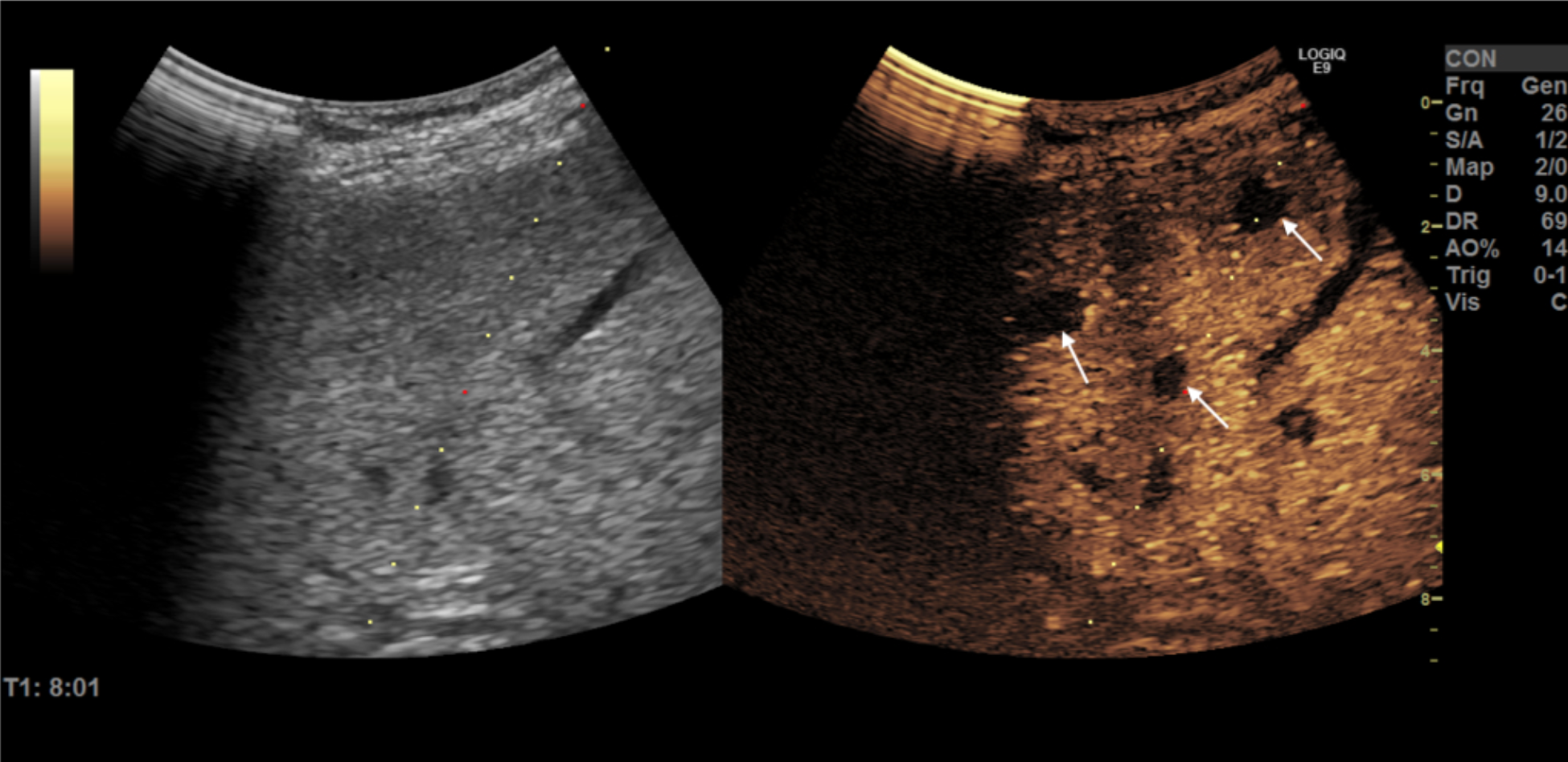
A 66-year-old male with a history of colon cancer was referred for US-guided biopsy to confirm small PET-positive liver-lesions suspicious of colorectal liver metastases. However, B-mode ultrasound imaging could not visualize the lesions why a Contrast-enhanced ultrasound (CEUS) study using Sonazoid was performed.
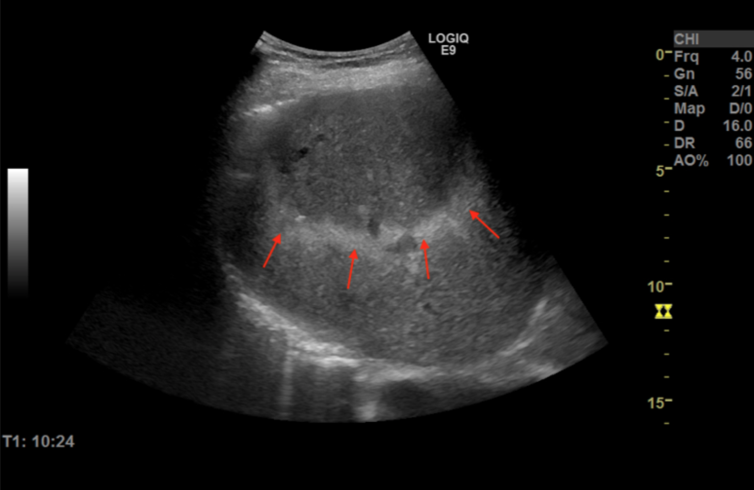
CEUS clearly demonstrated wash out of several small lesions, and a CEUS-guided biopsy was carried out at 8 minutes after contrast injection. Immediately after the procedure a strange phenomenon was seen on the B-mode image.
Quiz-summary
0 of 1 questions completed
Questions:
- 1
Information
View the March Case below, answer the question and then click check >
You have already completed the quiz before. Hence you can not start it again.
Quiz is loading...
You must sign in or sign up to start the quiz.
You have to finish following quiz, to start this quiz:
Results
0 of 1 questions answered correctly
Your time:
Time has elapsed
You have reached 0 of 0 points, (0)
Categories
- Not categorized 0%
- 1
- Answered
- Review
-
Question 1 of 1
1. Question
Question: What does the Northern Light phenomenon represent?
Correct
CORRECT ANSWER EXPLAINED BELOW Explanation
The Northern Lights phenomenon is caused by micro bubbles bursting during B-mode scanning at a point in time after Sonazoid administration when the organ in question (here the liver) is still packed with microbubbles.
The high density of Sonazoid bubbles will cause attenuation of sound, thus initially preventing sound propagation to deeper layers. At the same time it will burst the microbubbles due to the high MI inherent with B-mode imaging (as opposed to the deliberate setting of very low MI during CEUS scanning) thus generating a hyper-enhancing band horizontally across the image plane first superficially and later in the deeper parts.
The bursting bubbles are seen as a hyper-enhancing band propagating from the near filed towards deeper tissue layers when the most superficially placed bubbles are cleared by the first sound waves thus paving the way for the next wave – and the next and the next… and so forth still moving deeper down bursting its way through.
The artifact is well described and actually was part of the diagnostic procedure back in the early days when bubble disruption mode was part of the CEUS algorithm (1). It has been named “The gray-scale veil” to emphasize that this high MI CEUS artifact presents as a ‘Veil’ moving down into the deeper tissue parts as a veil covering underlying information (2)
References
- Skjoldbye B, Pedersen MH, Struckmann J, Burcharth F, Larsen T. Improved detection and biopsy of solid liver lesions using pulse-inversion ultrasound scanning and contrast agent infusion. Ultrasound Med Biol 2002;28:439-444.
- Wilson SR, Burns PN, Muradali D, Wilson JA, Lai X. Harmonic hepatic US with microbubble contrast agent: initial experience showing improved characterization of hemangioma, hepatocellular carcinoma, and metastasis. Radiology. 2000;215(1):153-61.
Incorrect
CORRECT ANSWER EXPLAINED BELOW Explanation
The Northern Lights phenomenon is caused by micro bubbles bursting during B-mode scanning at a point in time after Sonazoid administration when the organ in question (here the liver) is still packed with microbubbles.
The high density of Sonazoid bubbles will cause attenuation of sound, thus initially preventing sound propagation to deeper layers. At the same time it will burst the microbubbles due to the high MI inherent with B-mode imaging (as opposed to the deliberate setting of very low MI during CEUS scanning) thus generating a hyper-enhancing band horizontally across the image plane first superficially and later in the deeper parts.
The bursting bubbles are seen as a hyper-enhancing band propagating from the near filed towards deeper tissue layers when the most superficially placed bubbles are cleared by the first sound waves thus paving the way for the next wave – and the next and the next… and so forth still moving deeper down bursting its way through.
The artifact is well described and actually was part of the diagnostic procedure back in the early days when bubble disruption mode was part of the CEUS algorithm (1). It has been named “The gray-scale veil” to emphasize that this high MI CEUS artifact presents as a ‘Veil’ moving down into the deeper tissue parts as a veil covering underlying information (2)
References
- Skjoldbye B, Pedersen MH, Struckmann J, Burcharth F, Larsen T. Improved detection and biopsy of solid liver lesions using pulse-inversion ultrasound scanning and contrast agent infusion. Ultrasound Med Biol 2002;28:439-444.
- Wilson SR, Burns PN, Muradali D, Wilson JA, Lai X. Harmonic hepatic US with microbubble contrast agent: initial experience showing improved characterization of hemangioma, hepatocellular carcinoma, and metastasis. Radiology. 2000;215(1):153-61.