Quiz-summary
0 of 2 questions completed
Questions:
- 1
- 2
Information
You have already completed the quiz before. Hence you can not start it again.
Quiz is loading...
You must sign in or sign up to start the quiz.
You have to finish following quiz, to start this quiz:
Results
0 of 2 questions answered correctly
Time has elapsed
Categories
- Not categorized 0%
-
That is Case 08 completed – to move on to Case 09 ~ click here
- 1
- 2
- Answered
- Review
-
Question 1 of 2
1. Question
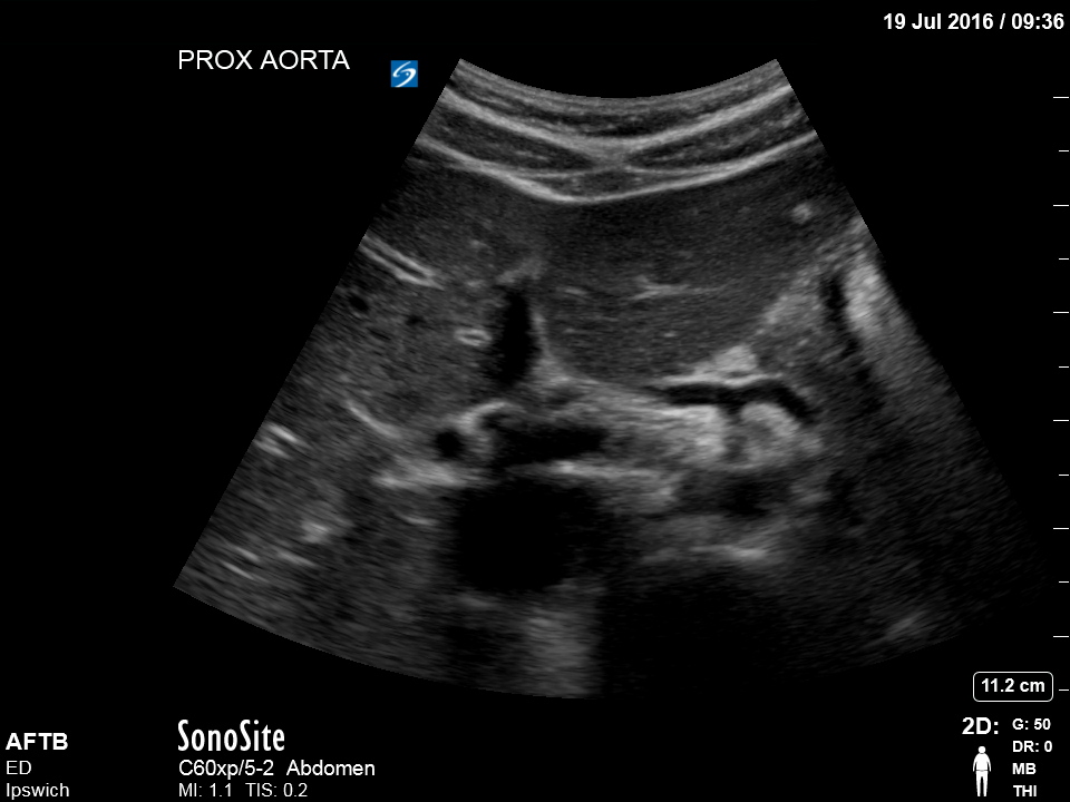
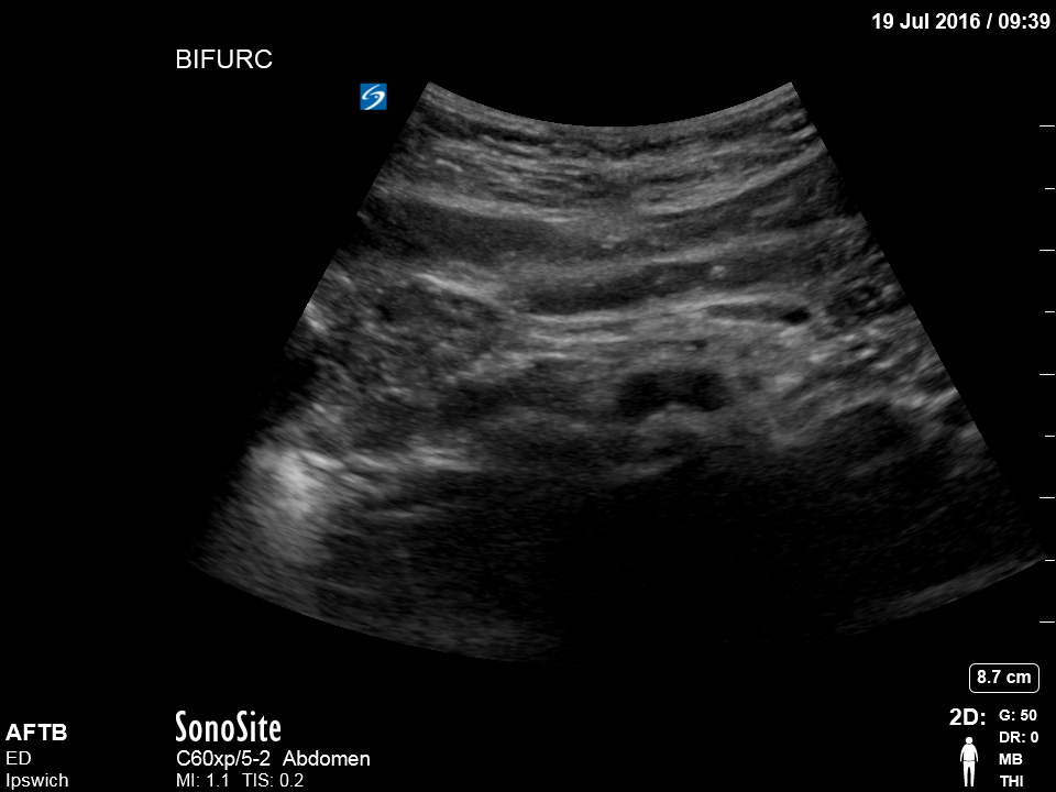
Clinical Description:
70 year old man presents with severe left sided back pain after playing a 3 day bowls tournament. He is an active man with no prior history of back pain or trauma. He has treated hypertension and no surgical history. His observations are normal other than a BP of 170/80.
Which of the following are true (NB there is more than 1 correct answer)
Correct
Incorrect
-
Question 2 of 2
2. Question
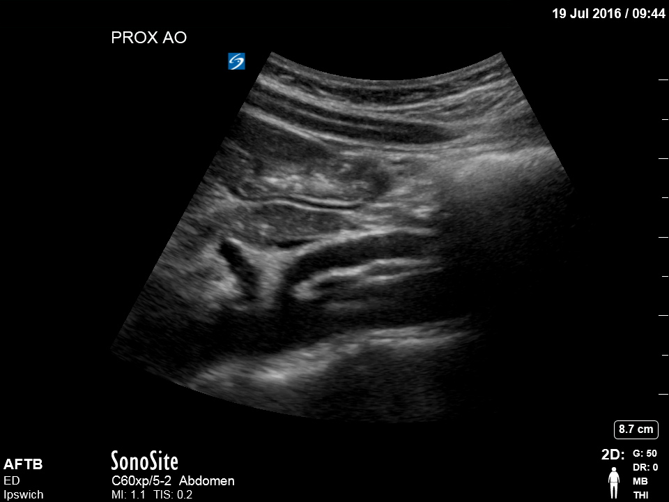
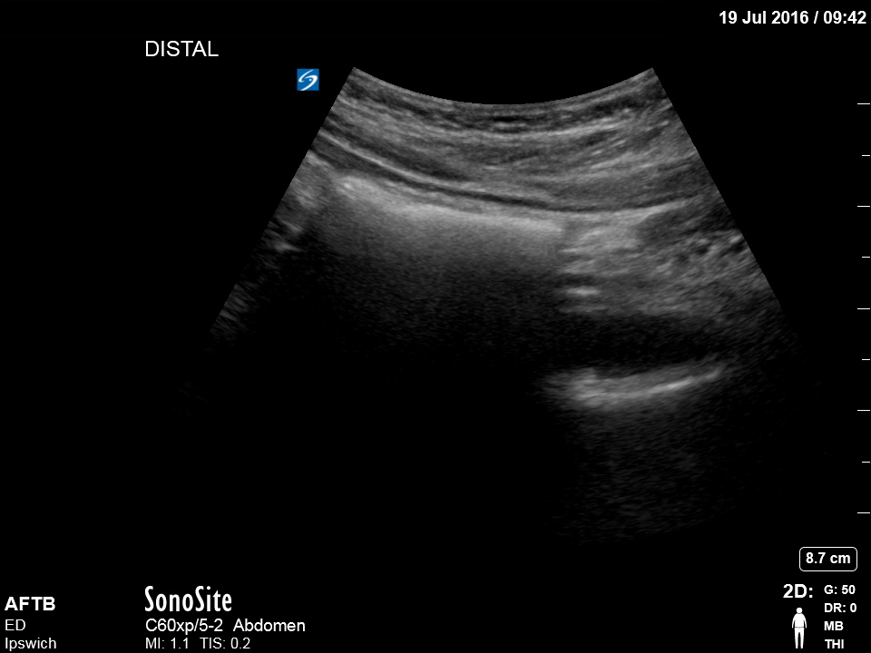
Clinical Description:
75 yr old male. Sudden onset epigastric and back pain. Shocked, diaphoretic, ashen
Regarding the use of Doppler, measurement and colour in this scan set, select the (one) best answer.
Correct
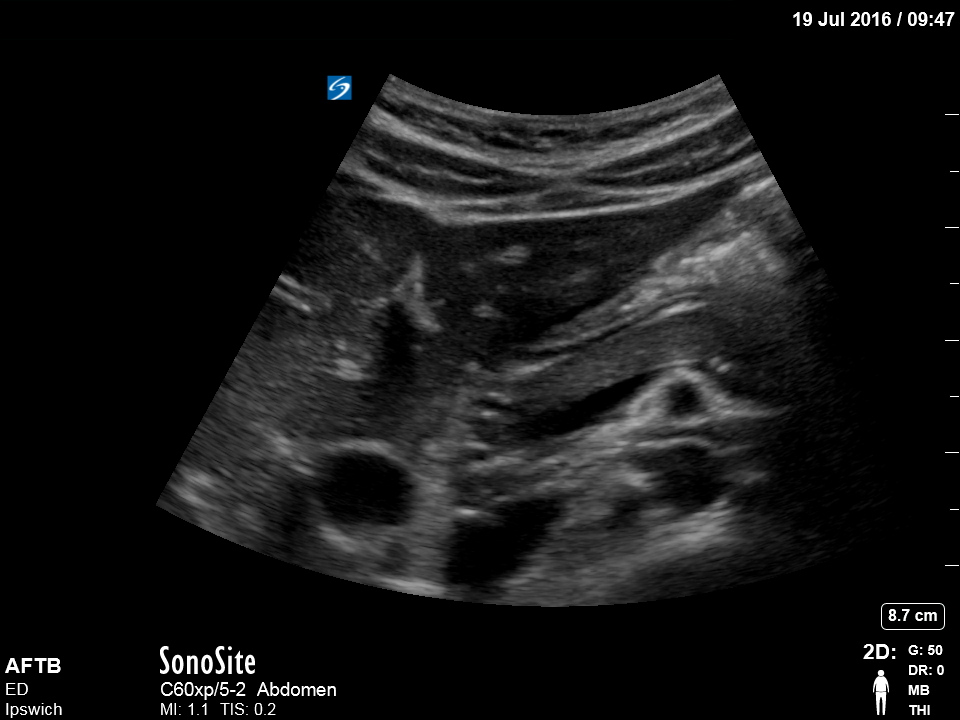
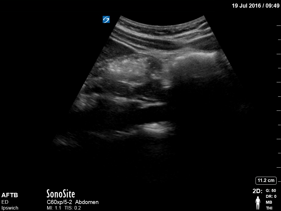
Lateral smearing increases with depth (as the beam widens). Transverse imaging with the probe tilted cranial or caudal will increase the apparent vertical diameter.
Both of these effects occur but will not change the assessment or management in the emergency or POCUS setting.– The spectral Doppler here is only used qualitatively, to show arterial pulse wave (proving the vessel is an artery – but not which artery). The Doppler angle is too great to allow any measurement of flow rate in this picture. There is very little need for either colour Doppler or spectral Doppler in POCUS evaluation of the aorta for AAA.
Incorrect